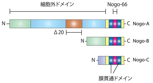
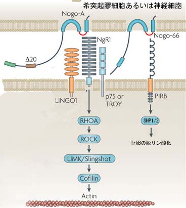
Two binding sites are currently known for the Nogo-66 sequence, the Nogo receptor 1 (NgR1) and the membrane protein paired immunoglobulin-like receptor B (PIRB). Both receptors also interact with other ligands, however. The receptor for the Nogo-A specific active site remains to be characterized. Rho activation followed by destabilizing effects on the cytoskeleton are obligatory steps in the postreceptor signalling and effector pathway that leads to the collapse of neurite growth cones. Several additional proteins are associated with what is probably a multisubunit receptor complex for Nogo-A.<br>Nogo-B, by interaction with a Nogo-B receptor (NGBR), influences vascular endothelial cells and smooth muscle cells, which hyperproliferate after vascular lesions in Nogo-A and Nogo-B double knockout mice. The function of Nogo-C is currently still unknown.<br>During CNS development, Nogo-A and its receptors are expressed in cortical precursors and affect their migration. Many projection neurons in the central and peripheral nervous systems express Nogo-A during axonal outgrowth; its neutralization or knockout enhances axonal fasciculation and influences branching. NgR1 and the shorter Nogo forms also have guidance and fasciculation functions in zebrafish, a lower vertebrate.<br>In the adult CNS, oligodendrocyte and myelin Nogo-A suppresses the growth programme of adult neurons, probably by a retrograde action on the cell bodies. Locally, neurite growth is dampened by the growth cone collapsing actions of Nogo-A. Nogo-A thus acts as a stabilizer of the adult CNS neuronal network and wiring. Ablation of Nogo-A or NgR1 accordingly enhances plastic rearrangements of CNS connections, extending the so-called 'critical period' far into adult ages, for example, for visual cortex plasticity. The schizophrenia-like behaviour of Nogo-A knockout mice and the associations found between psychiatric disorders and mutations in the genes encoding Nogo or NgR1 may be based on similar functions.<br>In addition to its cell surface expression, high amounts of Nogo are also present intracellularly. In neurons, its interaction with β-secretase points to a role in the regulation of amyloid precursor protein (APP) processing. Manipulations of Nogo have indicated a structural role for Nogo in the endoplasmic reticulum (ER) and the nuclear membrane. Interactions with proteins involved in cell survival and apoptosis have also been observed.<br>Various approaches aimed at suppressing Nogo-A or NgR1 actions have been used following injury of the adult spinal cord or brain. Acute functional suppression and, with more variable effects, chronic genetic deletion enhance regenerative sprouting and growth of various CNS tract systems. In addition, spared fibre systems have shown enhanced compensatory sprouting; both these processes were associated with substantial improvements of the behavioural recovery of lost functions in rodents and monkeys. These results illustrate the important growth-suppressive role of Nogo-A in the adult mammalian CNS.
Two binding sites are currently known for the Nogo-66 sequence, the Nogo receptor 1 (NgR1) and the membrane protein paired immunoglobulin-like receptor B (PIRB). Both receptors also interact with other ligands, however. The receptor for the Nogo-A specific active site remains to be characterized. Rho activation followed by destabilizing effects on the cytoskeleton are obligatory steps in the postreceptor signalling and effector pathway that leads to the collapse of neurite growth cones. Several additional proteins are associated with what is probably a multisubunit receptor complex for Nogo-A. Nogo-B, by interaction with a Nogo-B receptor (NGBR), influences vascular endothelial cells and smooth muscle cells, which hyperproliferate after vascular lesions in Nogo-A and Nogo-B double knockout mice. The function of Nogo-C is currently still unknown. During CNS development, Nogo-A and its receptors are expressed in cortical precursors and affect their migration. Many projection neurons in the central and peripheral nervous systems express Nogo-A during axonal outgrowth; its neutralization or knockout enhances axonal fasciculation and influences branching. NgR1 and the shorter Nogo forms also have guidance and fasciculation functions in zebrafish, a lower vertebrate. In the adult CNS, oligodendrocyte and myelin Nogo-A suppresses the growth programme of adult neurons, probably by a retrograde action on the cell bodies. Locally, neurite growth is dampened by the growth cone collapsing actions of Nogo-A. Nogo-A thus acts as a stabilizer of the adult CNS neuronal network and wiring. Ablation of Nogo-A or NgR1 accordingly enhances plastic rearrangements of CNS connections, extending the so-called 'critical period' far into adult ages, for example, for visual cortex plasticity. The schizophrenia-like behaviour of Nogo-A knockout mice and the associations found between psychiatric disorders and mutations in the genes encoding Nogo or NgR1 may be based on similar functions. In addition to its cell surface expression, high amounts of Nogo are also present intracellularly. In neurons, its interaction with β-secretase points to a role in the regulation of amyloid precursor protein (APP) processing. Manipulations of Nogo have indicated a structural role for Nogo in the endoplasmic reticulum (ER) and the nuclear membrane. Interactions with proteins involved in cell survival and apoptosis have also been observed. Various approaches aimed at suppressing Nogo-A or NgR1 actions have been used following injury of the adult spinal cord or brain. Acute functional suppression and, with more variable effects, chronic genetic deletion enhance regenerative sprouting and growth of various CNS tract systems. In addition, spared fibre systems have shown enhanced compensatory sprouting; both these processes were associated with substantial improvements of the behavioural recovery of lost functions in rodents and monkeys. These results illustrate the important growth-suppressive role of Nogo-A in the adult mammalian CNS.