テスト
ナビゲーションに移動
検索に移動
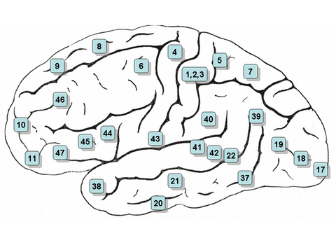
Image mapped Brodmann Areas. Clicking on an area in the picture causes the browser to load the appropriate article. 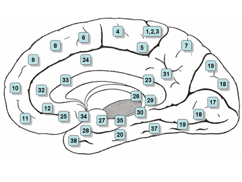
Image mapped Brodmann Areas. Clicking on an area in the picture causes the browser to load the appropriate article. Image mapped Brodmann Areas. Clicking on an area in the picture causes the browser to load the appropriate article. Image mapped Brodmann Areas. Clicking on an area in the picture causes the browser to load the appropriate article.
Brodmann areas for human & non-human primates
- ブロードマン3、1、2野 - 一次体性感覚野 (frequently referred to as Areas 3, 1, 2 by convention)
- ブロードマン4野 – 一次運動野
- ブロードマン5野 – 体性感覚連合野
- ブロードマン6野 - 前運動野 、補足運動野
- ブロードマン7野 -体性感覚連合野]
- ブロードマン8野 - 前頭眼野
- ブロードマン9野 - 前頭前野背外側部
- ブロードマン10野 - 前頭極
- ブロードマン11野 - 眼窩前頭野
- ブロードマン12野 - 眼窩前頭野
- ブロードマン13野 and ブロードマン14野* - 島皮質
- ブロードマン15野* - Anterior Temporal Lobe
- ブロードマン17野 - 一次視覚野 (V1)
- ブロードマン18野 - 二次視覚野(V2)]
- ブロードマン19野 - 視覚連合野(V3)
- ブロードマン20野 - 下側頭回
- ブロードマン21野 - 中側頭回
- ブロードマン22野 - 上側頭回
- ブロードマン23野 - 腹側後帯状皮質
- ブロードマン24野 - 腹側前帯状皮質
- ブロードマン25野 – 膝下野
- ブロードマン26野 - Ectosplenial portion of the retrosplenial region of the cerebral cortex
- ブロードマン27野 - 梨状葉皮質
- ブロードマン28野 - 後嗅内皮質
- ブロードマン29野 - 脳梁膨大後部帯状皮質
- ブロードマン30野 - 帯状皮質の一部
- ブロードマン31野 - 背側後帯状皮質
- ブロードマン32野 - 背側前帯状皮質
- ブロードマン33野 - 前帯状皮質の一部
- ブロードマン34野 - 前嗅内皮質 (on the 海馬傍回)
- ブロードマン35野 - 嗅周囲皮質 (on the 海馬傍回)
- ブロードマン36野 - 海馬傍回皮質 (on the 海馬傍回皮質)
- ブロードマン37野 - 紡錘状回
- ブロードマン38野 - 側頭極
- ブロードマン39野 - 角回
- ブロードマン40野 - 縁上回
- ブロードマン41、42野 - 一次聴覚野
- ブロードマン43野 - Primary gustatory cortex
- ブロードマン44野 - 下前頭回弁蓋部
- ブロードマン45野 - 下前頭回 三角部
- ブロードマン46野 - 前頭前野背外側部
- ブロードマン47野 - 下前頭前野
- ブロードマン48野 - Retrosubicular area (a small part of the medial surface of the temporal lobe)
- ブロードマン49野 - Parasubiculum area in a rodent
- ブロードマン52野 - Parainsular area (at the junction of the temporal lobe and the insula)
(*) Area only found in non-human primates.
Some of the original Brodmann areas have been subdivided further, e.g., "23a" and "23b".[1]
Clickable map: Lateral Surface
Clickable map: Medial Surface
- ↑ Brent A. Vogt, Deepak N. Pandya, Douglas L. Rosene (1987). "Cingulate cortex of the rhesus monkey: I. Cytoarchitecture and thalamic afferents". The Journal of Comparative Neurology. 262 (2): 256–270. doi:10.1002/cne.902620207. PMID 3624554.
{{cite journal}}: Unknown parameter|month=ignored (help)CS1 maint: multiple names: authors list (link)
Brodmann areas for human & non-human primates
- Areas 3, 1 & 2 - Primary Somatosensory Cortex (frequently referred to as Areas 3, 1, 2 by convention)
- Area 4 - Primary Motor Cortex
- Area 5 - Somatosensory Association Cortex
- Area 6 - Premotor cortex and Supplementary Motor Cortex (Secondary Motor Cortex)(Supplementary motor area)
- Area 7 - Somatosensory Association Cortex
- Area 8 - Includes Frontal eye fields
- Area 9 - Dorsolateral prefrontal cortex
- Area 10 - Anterior prefrontal cortex (most rostral part of superior and middle frontal gyri)
- Area 11 - Orbitofrontal area (orbital and rectus gyri, plus part of the rostral part of the superior frontal gyrus)
- Area 12 - Orbitofrontal area (used to be part of BA11, refers to the area between the superior frontal gyrus and the inferior rostral sulcus)
- Area 13 and Area 14* - Insular cortex
- Area 15* - Anterior Temporal Lobe
- Area 17 - Primary visual cortex (V1)
- Area 18 - Secondary visual cortex (V2)
- Area 19 - Associative visual cortex (V3,V4,V5)
- Area 20 - Inferior temporal gyrus
- Area 21 - Middle temporal gyrus
- Area 22 - Superior temporal gyrus, of which the caudal part is usually considered to contain the Wernicke's area
- Area 23 - Ventral Posterior cingulate cortex
- Area 24 - Ventral Anterior cingulate cortex.
- Area 25 - Subgenual cortex (part of the Ventromedial prefrontal cortex)[1]
- Area 26 - Ectosplenial portion of the retrosplenial region of the cerebral cortex
- Area 27 - Piriform cortex
- Area 28 - Posterior Entorhinal Cortex
- Area 29 - Retrosplenial cingulate cortex
- Area 30 - Part of cingulate cortex
- Area 31 - Dorsal Posterior cingulate cortex
- Area 32 - Dorsal anterior cingulate cortex
- Area 33 - Part of anterior cingulate cortex
- Area 34 - Anterior Entorhinal Cortex (on the Parahippocampal gyrus)
- Area 35 - Perirhinal cortex (on the Parahippocampal gyrus)
- Area 36 - Parahippocampal cortex (on the Parahippocampal gyrus)
- Area 37 - Fusiform gyrus
- Area 38 - Temporopolar area (most rostral part of the superior and middle temporal gyri)
- Area 39 - Angular gyrus, considered by some to be part of Wernicke's area
- Area 40 - Supramarginal gyrus considered by some to be part of Wernicke's area
- Areas 41 & 42 - Primary and Auditory Association Cortex
- Area 43 - Primary gustatory cortex
- Area 44 - pars opercularis, part of Broca's area
- Area 45 - pars triangularis Broca's area
- Area 46 - Dorsolateral prefrontal cortex
- Area 47 - Inferior prefontal gyrus
- Area 48 - Retrosubicular area (a small part of the medial surface of the temporal lobe)
- Area 49 - Parasubiculum area in a rodent
- Area 52 - Parainsular area (at the junction of the temporal lobe and the insula)
(*) Area only found in non-human primates.
Some of the original Brodmann areas have been subdivided further, e.g., "23a" and "23b".[2]
Clickable map: Lateral Surface
Clickable map: Medial Surface
- ↑ http://ukpmc.ac.uk/articles/PMC2268639;jsessionid=BBF4DB8DAFCFCB452BEA9AB7368AB5C6.jvm4
- ↑ Brent A. Vogt, Deepak N. Pandya, Douglas L. Rosene (1987). "Cingulate cortex of the rhesus monkey: I. Cytoarchitecture and thalamic afferents". The Journal of Comparative Neurology. 262 (2): 256–270. doi:10.1002/cne.902620207. PMID 3624554.
{{cite journal}}: Unknown parameter|month=ignored (help)CS1 maint: multiple names: authors list (link)