WikiSysopによるアップロード
ナビゲーションに移動
検索に移動
この特別ページでは、アップロードされたファイルをすべて表示します。
| 日時 | 名前 | サムネイル | サイズ | 概要 | 版数 |
|---|---|---|---|---|---|
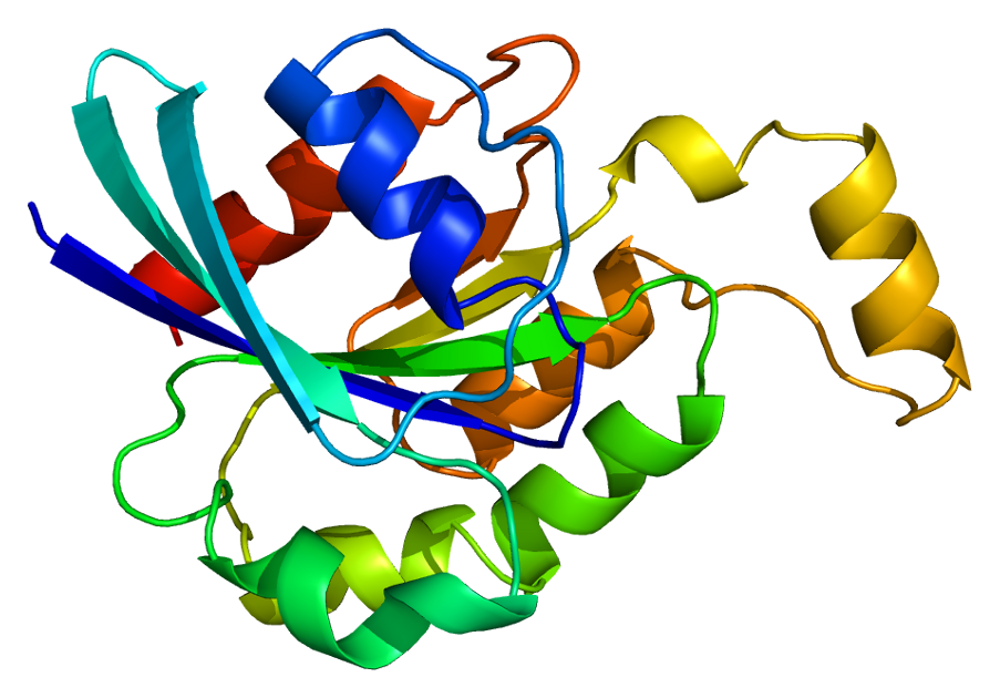
| 2013年1月13日 (日) 13:16 | Protein MEF2A PDB 1c7u.png (ファイル) |  |
303キロバイト | == {{int:filedesc}} == {{Information | Description={{en | 1=Structure of the MEF2A protein. Based on PyMOL rendering of PDB [http://www.pdb.org/pdb/explore/explore.do?structureId=1c7u 1c7u].}} | Source={{own}} | Author=Emw | 1 |
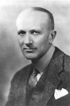
| 2012年12月8日 (土) 23:18 | Wilder Penfield.jpg (ファイル) |  |
17キロバイト | {{Information |Description={{en|Wilder Penfield Source: [http://www.archives.mcgill.ca/pictures/pr000632.GIF McGill University Archives] PORTRAIT: WILDER PENFIELD, DIRECTOR OF THE MONTREAL NEUROLOGICAL INSTITUTE, 1938-1960 Date: 1934CA Size: 003.5 X 0 | 1 |
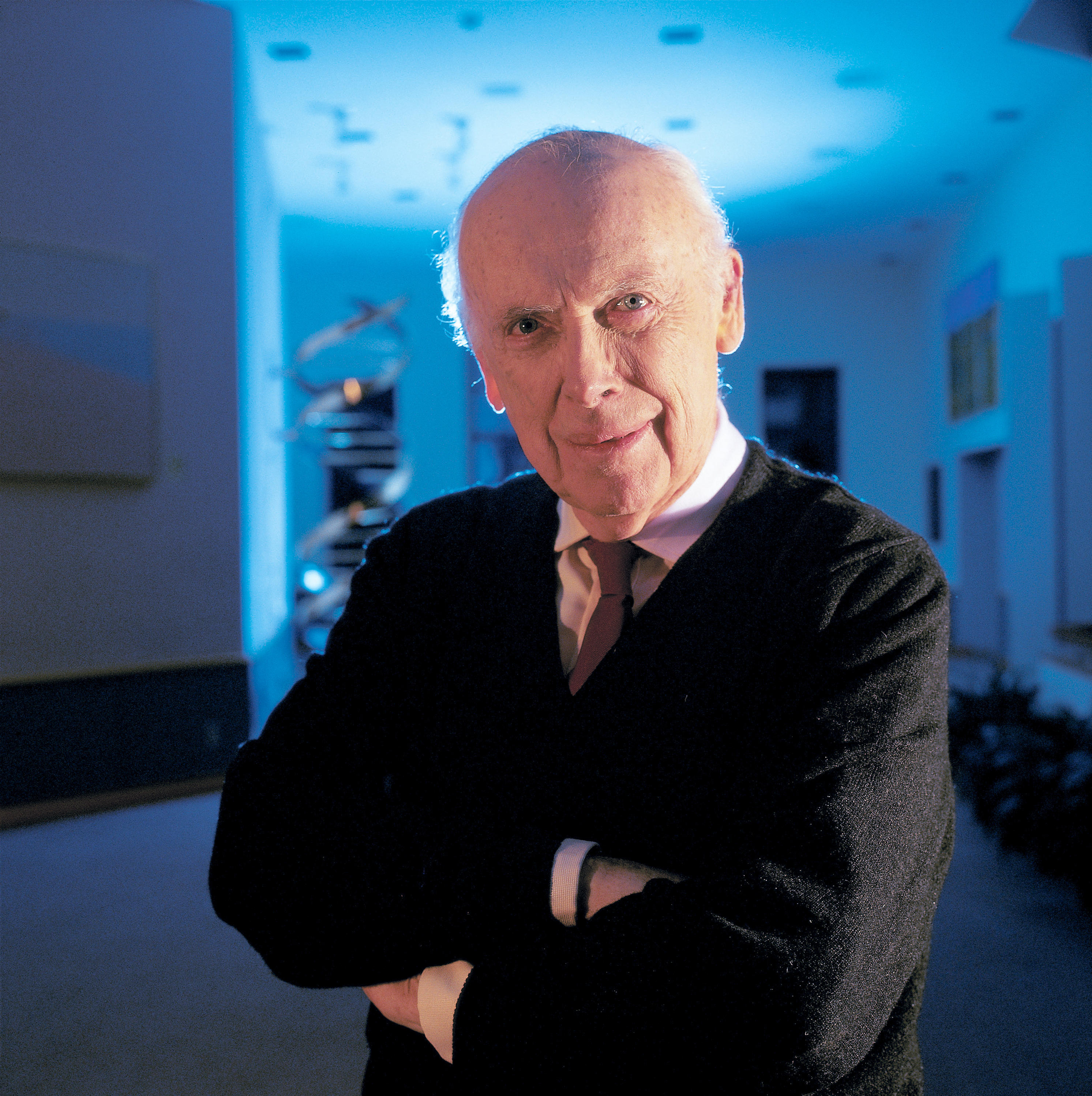
| 2012年12月8日 (土) 22:44 | James D Watson.jpg (ファイル) |  |
9.11メガバイト | == {{int:filedesc}} == {{Information |Description={{en|1=Nobel laureate Dr. James D. Watson, Chancellor, Cold Spring Harbor Laboratory. These images are freely available and may be used without special permission.}} |Source={{Derived from|James_D_Watson_G | 1 |
| 2012年12月8日 (土) 18:41 | Brodmann Cytoarchitectonics.PNG (ファイル) |  |
173キロバイト | == {{int:filedesc}} == {{Information |Description={{en|1=Cytoarchitectonics of human brain according to Brodmann (1909)}} |Source=Vergleichende Lokalisationslehre der Grosshirnrinde in ihren Prinzipien dargestellt auf Grund des Zellenbaues, Johann Ambrosi | 1 |
| 2012年12月6日 (木) 00:49 | Brain diagram without text.svg (ファイル) |  |
40キロバイト | == {{int:filedesc}} == {{Information |Description={{en|Principal lobes of the cerebrum viewed laterally. Figure 728 from Gray's Anatomy. 4 lines note sulci as follows *top center: Central sulcus *top right: Parieto-occipital sulcus *down left: Lateral sul | 1 |
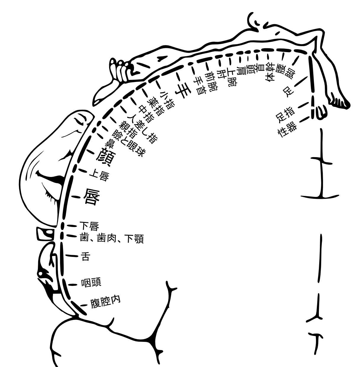
| 2012年12月4日 (火) 09:27 | Somatosensory cortex ja.png (ファイル) |  |
209キロバイト | {{Information| |Description = diagram showing position of regions of the human cortex corresponding to the respective afferent nerve region of the body. Writing in Japanese language. 感覚皮質における身体の対応部位を示した図。ペンフ | 1 |
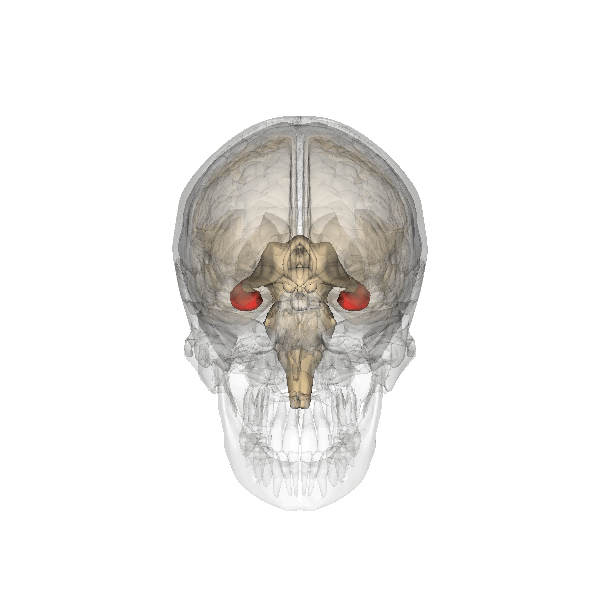
| 2012年11月29日 (木) 00:41 | Hippocampus.gif (ファイル) |  |
4.09メガバイト | {{Information |Description={{en|1=hippocampus. Images are from Anatomography maintained by Life Science Databases(LSDB). }} {{ja|1=海馬。 画像はLife Science Databases(LSDB)のAnatomographyというサイトより。}} |Source=from Anatomography, we | 1 |
| 2012年11月24日 (土) 18:07 | Cc.logo.circle.svg (ファイル) | 1キロバイト | 1 | ||
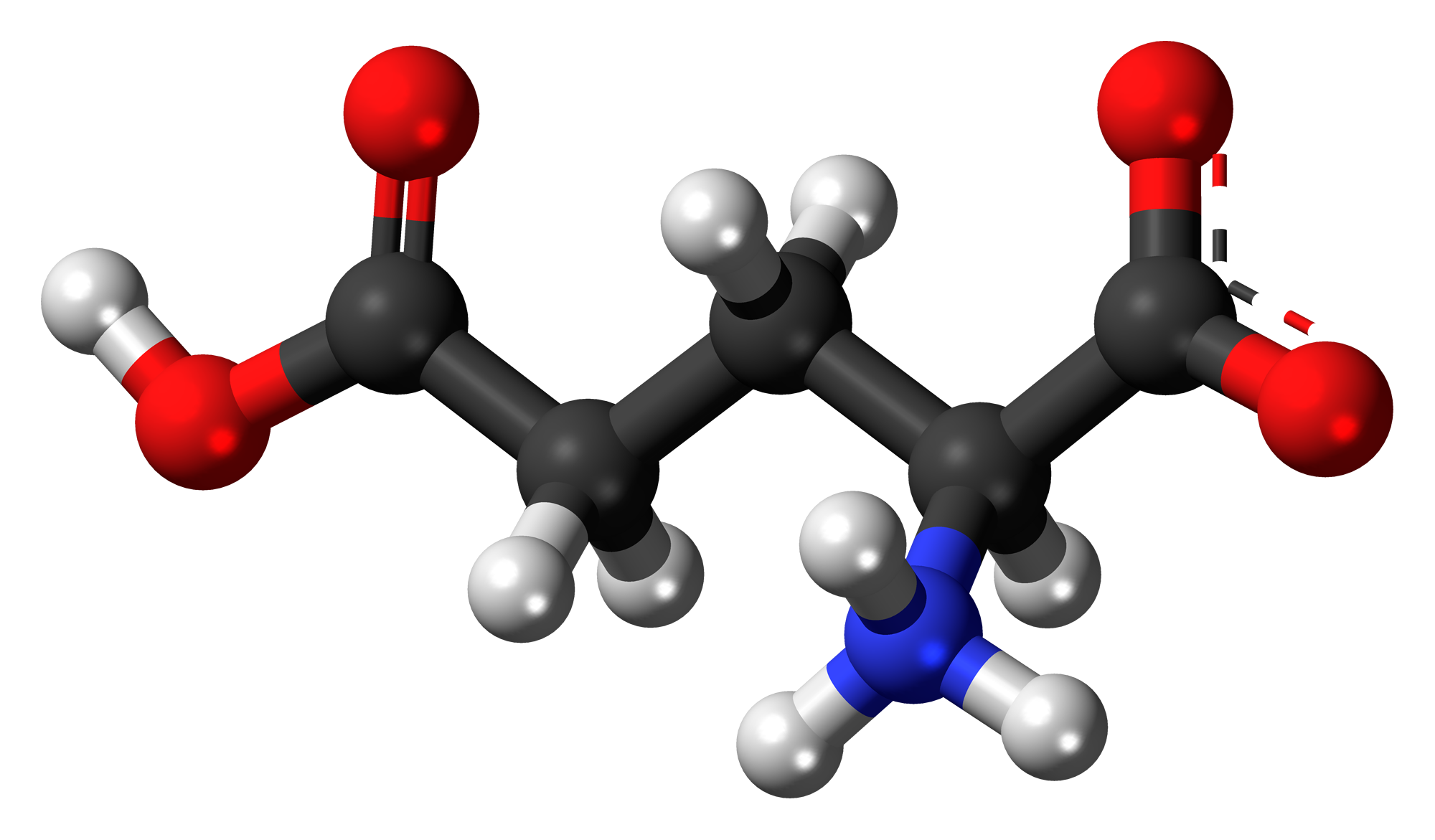
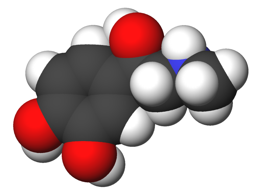
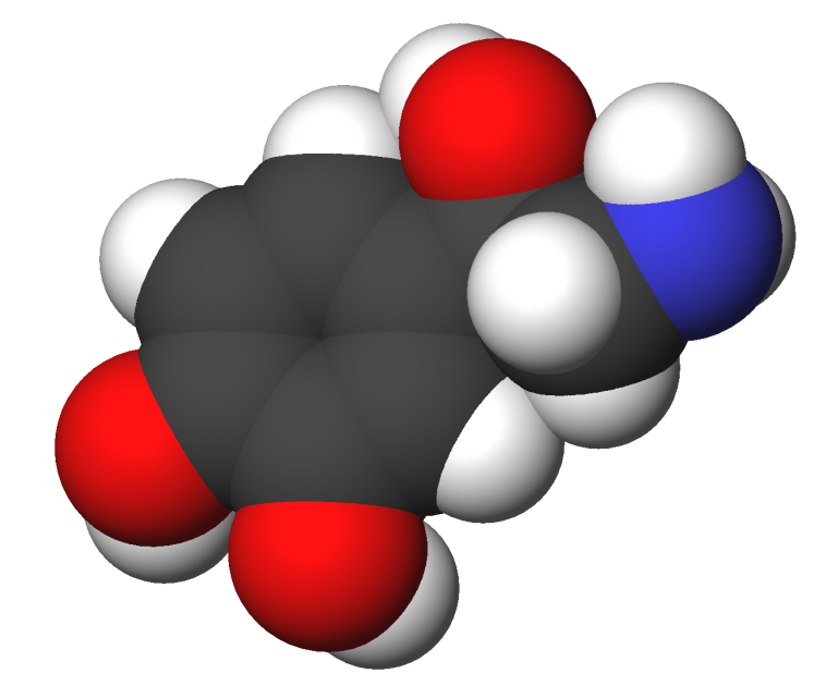
| 2012年11月24日 (土) 18:04 | L-Glutamic-acid-zwitterion-3D-balls.png (ファイル) |  |
362キロバイト | =={{int:filedesc}}== {{Information |description=Ball-and-stick model of the '''glutamic acid''' molecule, one of the 20 amino acids used to build proteins. This image shows the L isomer in [[w:zwitterion| | 1 |
| 2012年11月24日 (土) 17:59 | Glutaminsäure - Glutamic acid.svg (ファイル) |  |
9キロバイト | {{Information |Description=de: Struktur von Glutaminsäure;<br />en: Structure of glutamic acid |Source={{own}} |Date=2007-02-04 |Author=NEUROtiker |Permission=Own work, all rights released (Public domain) |other_versions= }} {{PD-sel | 1 |
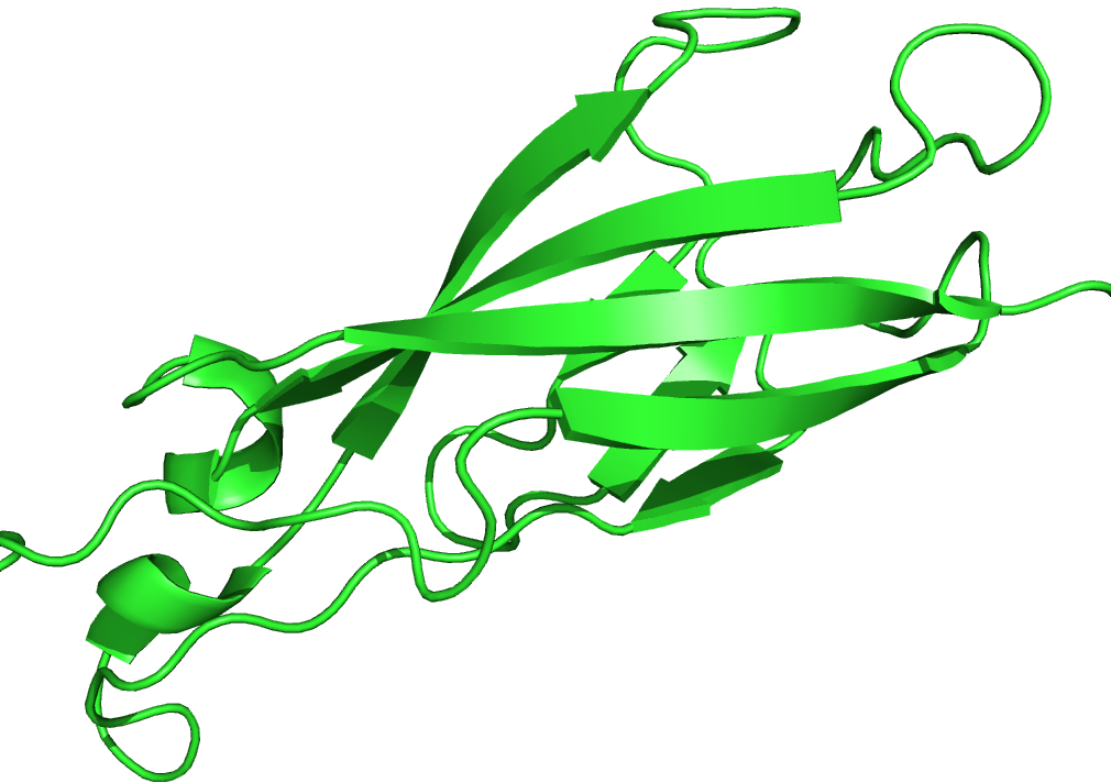
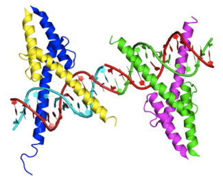
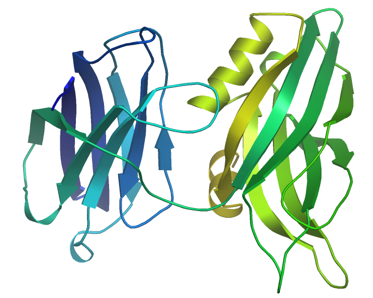
| 2012年11月1日 (木) 09:45 | 1mdm Pax5 paired domain.png (ファイル) |  |
176キロバイト | 1 | |
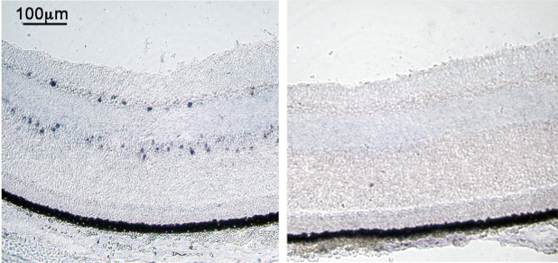
| 2012年10月28日 (日) 10:04 | In situハイブリダイゼーション法1D.png (ファイル) |  |
206キロバイト | 3 | |
| 2012年10月28日 (日) 10:04 | In situハイブリダイゼーション法1C.png (ファイル) |  |
522キロバイト | 3 | |
| 2012年10月28日 (日) 10:03 | In situハイブリダイゼーション法1B.png (ファイル) |  |
346キロバイト | 3 | |
| 2012年10月28日 (日) 10:02 | In situハイブリダイゼーション法1A.png (ファイル) |  |
426キロバイト | 3 | |
| 2012年10月28日 (日) 09:53 | In situハイブリダイゼーション法3.png (ファイル) | 47キロバイト | 2 | ||
| 2012年9月30日 (日) 00:44 | Epinephrine-3d-CPK.png (ファイル) |  |
105キロバイト | 1 | |
| 2012年9月29日 (土) 21:24 | Norepinephrine-3d-CPK.png (ファイル) |  |
96キロバイト | 1 | |
| 2012年9月27日 (木) 00:25 | Protein RHOA PDB 1a2b.png (ファイル) |  |
306キロバイト | 1 | |
| 2012年9月17日 (月) 00:13 | 1QLX.png (ファイル) |  |
91キロバイト | PDB entry 1qlx: HUMAN PRION PROTEIN | 1 |
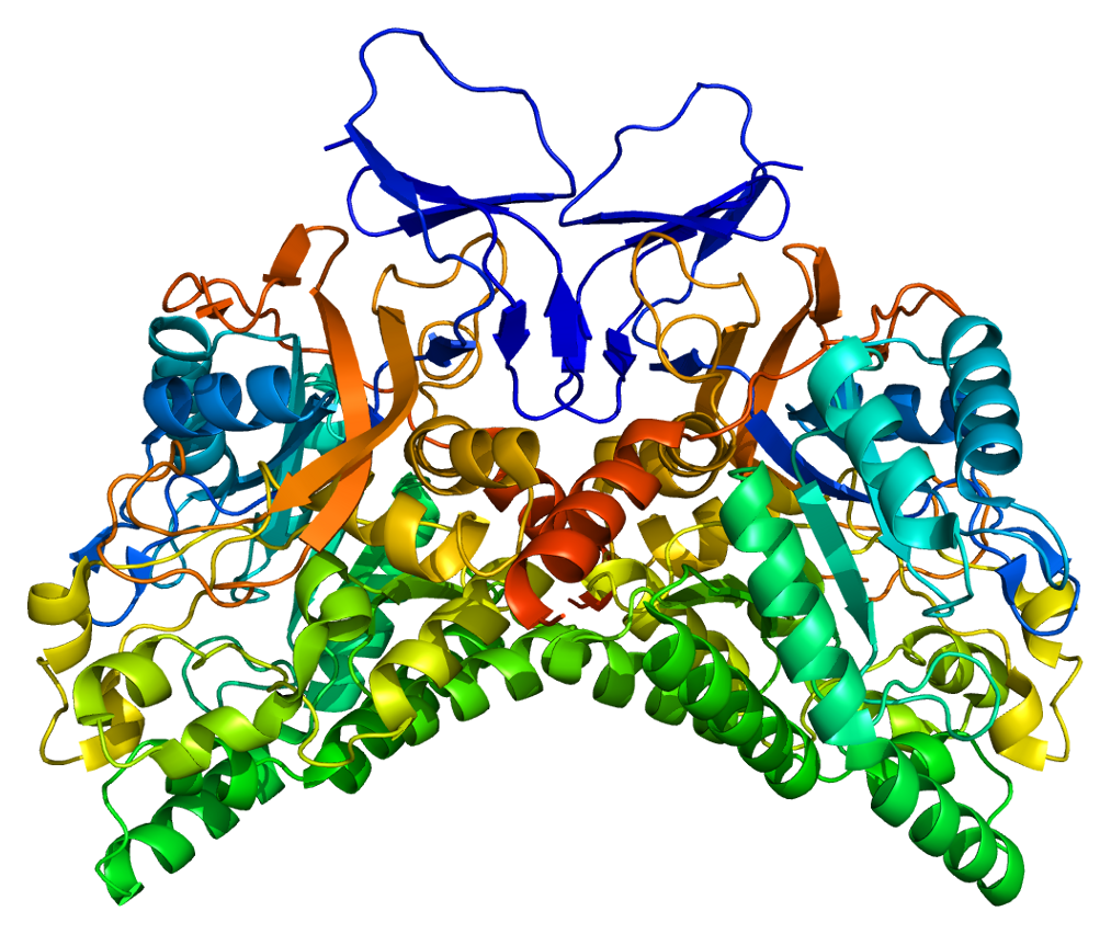
| 2012年9月6日 (木) 23:41 | ECadherin repeating unit.png (ファイル) |  |
181キロバイト | 1 | |
| 2012年9月2日 (日) 02:22 | Protein CRMP1 PDB 1kcx.png (ファイル) |  |
665キロバイト | 1 | |
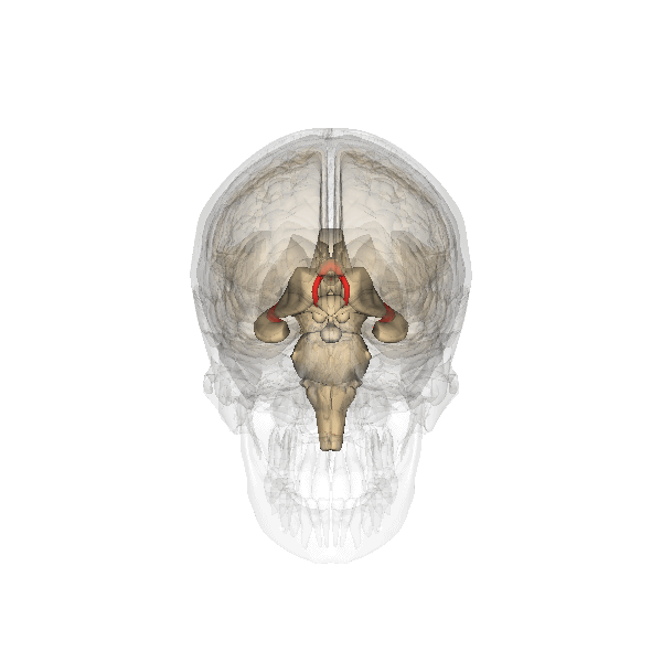
| 2012年8月20日 (月) 09:11 | Hypothalamus.gif (ファイル) |  |
2.03メガバイト | 1 | |
| 2012年8月19日 (日) 19:51 | Inferior colliculus.gif (ファイル) |  |
2.2メガバイト | 1 | |
| 2012年8月18日 (土) 20:49 | Gray747.png (ファイル) |  |
17キロバイト | 1 | |
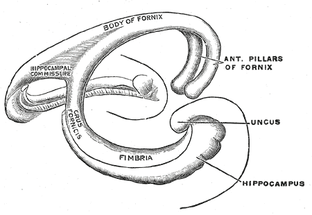
| 2012年8月18日 (土) 20:42 | Fornix.gif (ファイル) |  |
3.94メガバイト | 1 | |
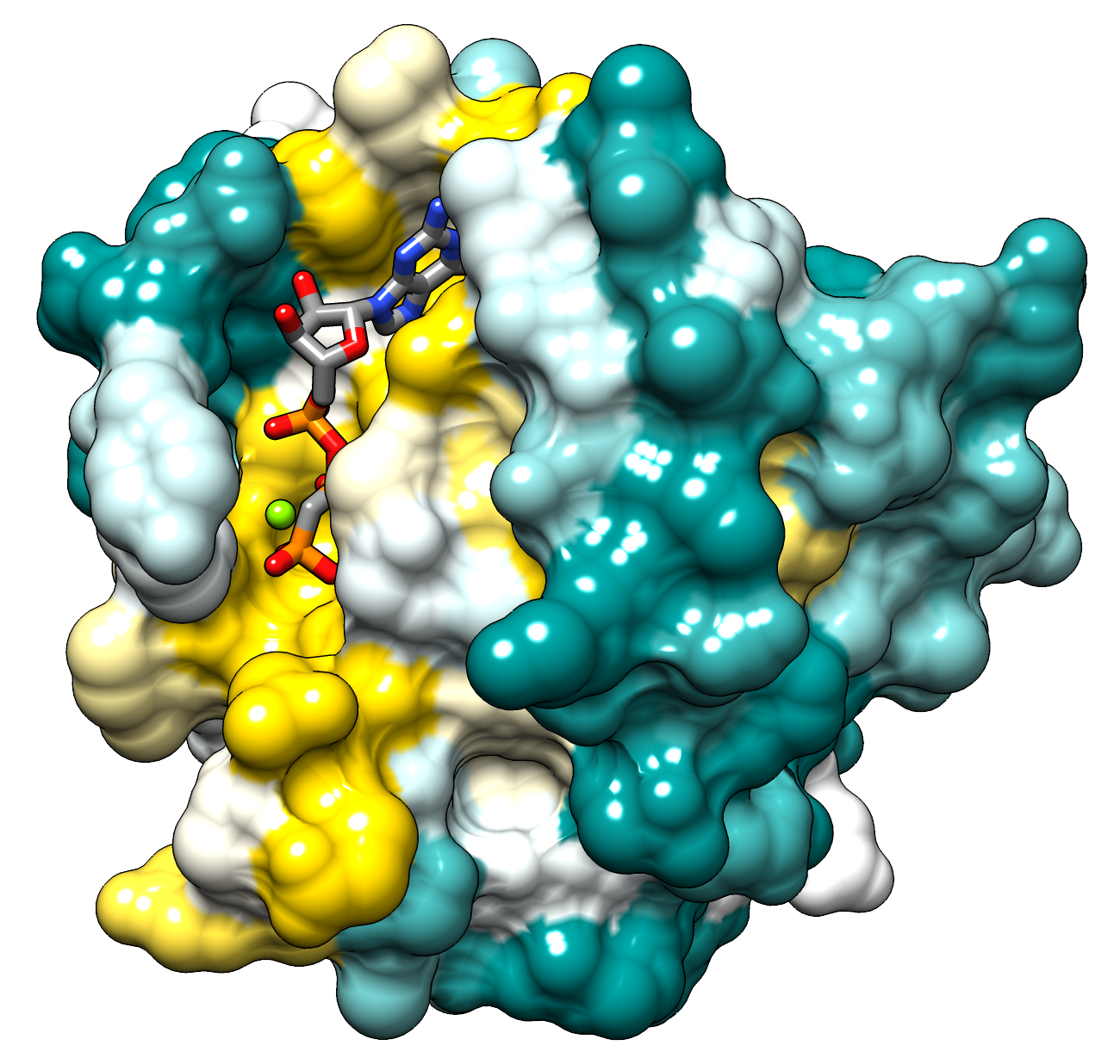
| 2012年7月26日 (木) 20:53 | Hras surface colored by conservation.png (ファイル) |  |
973キロバイト | 1 | |
| 2012年7月18日 (水) 23:37 | Toshiyukiotsuka pfambox.png (ファイル) |  |
77キロバイト | 1 | |
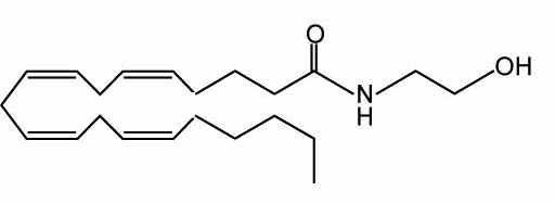
| 2012年7月18日 (水) 17:50 | Yukihashimotodani fig 1 anandamide.png (ファイル) | 10キロバイト | 1 | ||
| 2012年7月18日 (水) 17:46 | Yukihashimotodani fig 1 anadamide.png (ファイル) | 10キロバイト | 1 | ||
| 2012年7月18日 (水) 17:46 | Yukihashimotodani fig 1 2-AG.png (ファイル) | 12キロバイト | 1 | ||
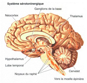
| 2012年7月16日 (月) 12:59 | Serotonergic system.png (ファイル) |  |
90キロバイト | 1 | |
| 2012年7月14日 (土) 00:18 | Huntington.jpg (ファイル) |  |
80キロバイト | 1 | |
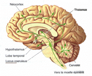
| 2012年7月13日 (金) 23:55 | Locus-coeruleus.png (ファイル) |  |
82キロバイト | 1 | |
| 2012年7月11日 (水) 22:52 | X mark.svg (ファイル) |  |
4キロバイト | 1 | |
| 2012年7月8日 (日) 13:48 | Protein BMP4 PDB 1reu.png (ファイル) |  |
141キロバイト | 1 | |
| 2012年6月28日 (木) 18:21 | Dammy 1pxX1px.png (ファイル) | 948バイト | 3 | ||
| 2012年6月15日 (金) 23:34 | Nagetaishihara fig 3.jpg (ファイル) |  |
109キロバイト | 2012年1月18日 (水)08:55の版へ差し戻し | 1 |
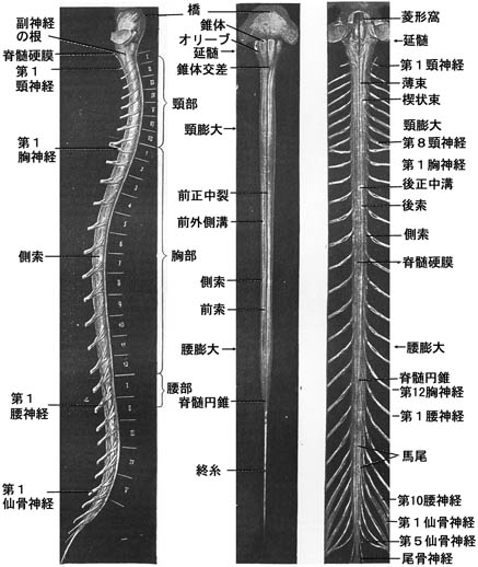
| 2012年5月29日 (火) 23:17 | Spinal cord with spinal nerve.jpg (ファイル) |  |
19キロバイト | 1 | |
| 2012年5月29日 (火) 23:16 | Spinal cord whole view.jpg (ファイル) |  |
54キロバイト | 1 | |
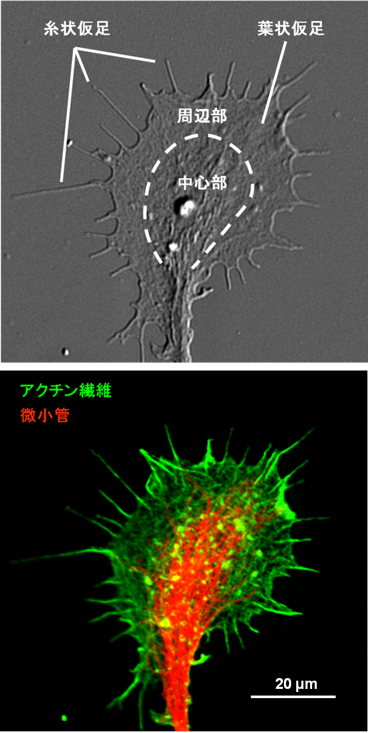
| 2012年5月27日 (日) 01:04 | 成長円錐拡大.png (ファイル) |  |
670キロバイト | 2 | |
| 2012年5月27日 (日) 01:02 | 成長円錐全体.png (ファイル) |  |
412キロバイト | 2 | |
| 2012年5月25日 (金) 16:56 | Tetonoff図1.png (ファイル) |  |
106キロバイト | 1 | |
| 2012年5月25日 (金) 16:56 | Tetonoff図2.png (ファイル) |  |
105キロバイト | 1 | |
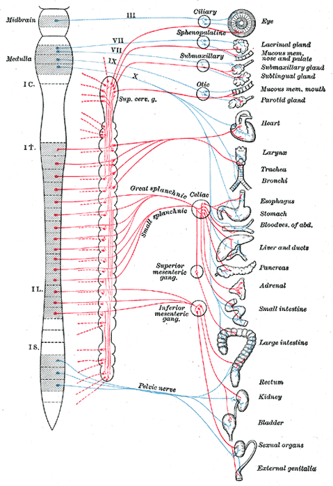
| 2012年5月23日 (水) 00:41 | Sympathetic nervous system.png (ファイル) |  |
166キロバイト | From Gray's anatomy | 1 |
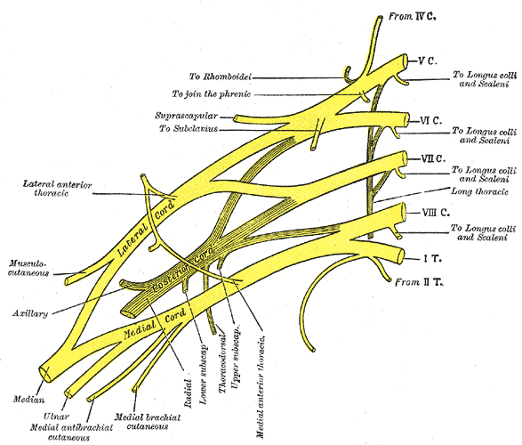
| 2012年5月23日 (水) 00:23 | Brachial plexus.png (ファイル) |  |
113キロバイト | From Gray's anatomy | 1 |
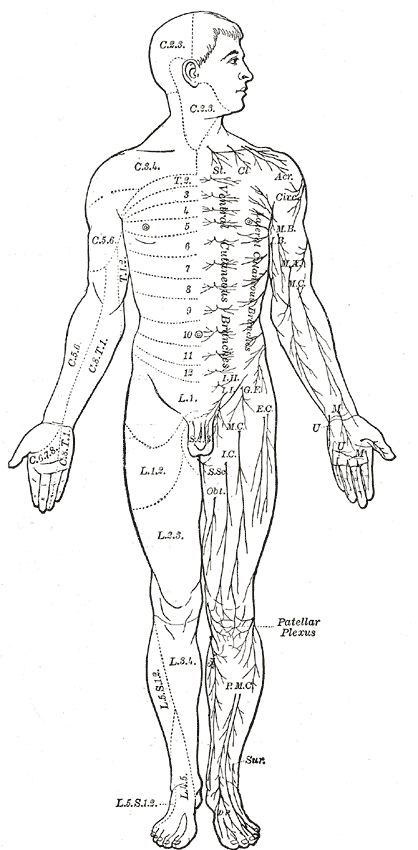
| 2012年5月23日 (水) 00:11 | Dermatome.png (ファイル) |  |
108キロバイト | From Gray's anatomy | 1 |
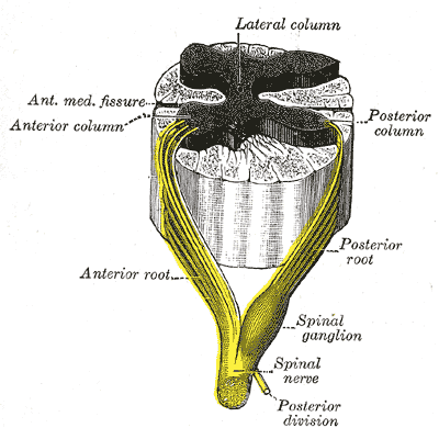
| 2012年5月23日 (水) 00:02 | Spinal cord.png (ファイル) |  |
69キロバイト | From Gray's anatomy | 1 |
| 2012年5月18日 (金) 18:15 | Synaptotagmin C2 domain.png (ファイル) |  |
164キロバイト | Crystal structure of Human Synaptotagmin 1 C2 domain. Drawn with Astex MolecularViewer based on 2R83 by user:yhayashi. | 1 |
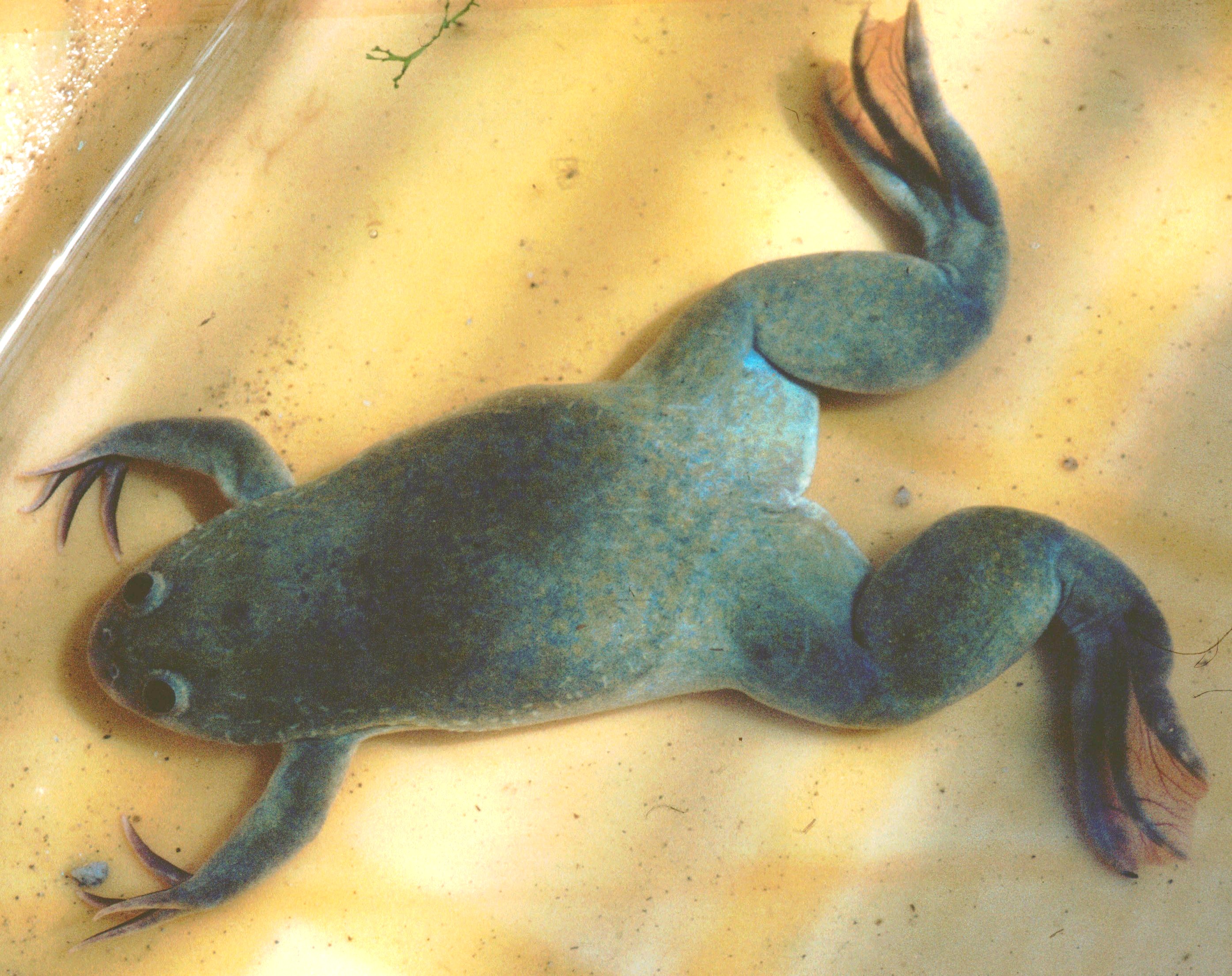
| 2012年5月15日 (火) 19:52 | Xenopus laevis 1.jpg (ファイル) |  |
1.34メガバイト | == {{int:filedesc}} == {{Information |Description=''Xenopus laevis'' |Source=de:wp ([http://de.wikipedia.org/wiki/Bild:Xenopus_laevis_1.jpg original image page here]) |Date=2005-11-04 |Author=de:User:Michael Linnenbach |other_versions= }} == {{int:l | 1 |