視覚前野
伊藤南
東京医科歯科大学生体機能支援システム学分野
DOI:10.14931/bsd.3705 原稿受付日:2013年5月24日 原稿完成日:2015年月日
担当編集委員:藤田一郎(大阪大学 大学院生命機能研究科)
英語名:extrastriate cortex、circumstriate cortex 独:extrastriärer Kortex 仏:cortex extrastrié
同義語:外線条皮質、有線外皮質、後頭連合野
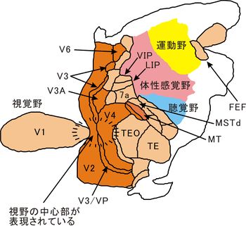
視覚前野(しかくぜんや)は哺乳類の大脳新皮質の視覚野の一部で、後頭葉の視覚連合野(後頭連合野)、ブロードマンの18、19野に相当する。さらにV2、V3、V3A、V4、V5/MT、V6等の機能的領野に区分される。第一次視覚野(V1、17野)より主な入力を受けて視覚情報処理を行う。各領野のニューロンは受容野を持ち、レチノトピーの性質を示して、片半球の領野が反対側の半視野を表す。これらの領野は階層的な結合関係を持ち、上の階層の領野ほど受容野が大きく、より複雑な刺激特徴や大局的な情報を抽出表現する。主に2つの視覚経路に分かれており、腹側視覚路はV2、V4を介して下側頭葉(側頭連合野)に出力し、物体の形状や物体表面の性質(明るさ、色、模様)を表し、視覚対象の認識や形状の表象に寄与する。背側視覚路はV2、V3、V5/MT、V6を介して後頭頂葉(頭頂連合野)に出力し、3次元的な空間配置、空間の構造、動きを表して、眼や腕の運動制御に寄与する。
視覚前野とは
哺乳類の大脳新皮質の視覚野の一部で、後頭葉の視覚連合野(後頭連合野)、あるいは後頭葉から一次視覚野(V1)を除いた部分。細胞構築学的にはブロードマンの脳地図の18野、19野に相当する。18野を前有線皮質(傍有線野、prestriate cortex)、19野を周有線皮質(周線条野、後頭眼野、parastriate cortex)、視覚前野全体を外線条皮質(有線外皮質、extrastriate cortex、circumstriate cortex)と呼ぶ。当初、一次視覚野(V1)に隣接する領域を広く視覚前野ないし視覚連合野と称した。1960年代以降、ニューロンの応答特性、受容野の大きさや位置、ニューロン間の結合関係に着目した領野区分の研究がネコやサルで盛んになった。また免疫組織化学的な染色法による細胞構築学的な研究も進んだ。1980年代以降、fMRIや光計測等のイメージング技術の発達により視野地図の広がりを可視化する研究が進んだ。現在ではV2、V3、V4、V5/MT、V6等の機能的な領野が同定され、個別の領野として扱われることが多い。機能的な領野区分は旧世界ザルのマカカ属サル(アカゲザル、ニホンザルなど)で最も進んでいるが、細部や高次領域(V3、V4、V6)については研究者間で見解の相違がある。動物種によっても区分法や名称が異なる。
機能的な領野の区分

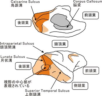
大脳皮質の表面をのばして表示したもので、内側で切って上下に開いたように表示してある。右側が前頭葉(前側)、左側が後頭葉(後側)。橙色の部分が視覚前野、肌色がその他の視覚野を示す。(Felleman and Van Essen (1991)[1] Fig.2を改変)
V1と同様に、視覚前野のニューロンは(古典的)受容野内に呈示された視覚刺激が持つ物理的な特性を抽出する。機能的な領野ごとに抽出される刺激特性が異なり、刺激特徴やそのパラメータに対する選択的な反応により表される。一方、視覚刺激の位置情報は受容野の位置で表される。各領野はレチノトピー(網膜部位の再現)の性質を示し(詳細は受容野を参照)、片半球の1つの領野が反対側の視野を映す一枚のトポグラフィックな視野地図を表す。受容野の位置が中心視野(fovea)から周辺視野に移るにつれて、受容野の大きさは一定の割合で大きくなる。マカカ属サルのV2、V3、V4はそれぞれV1の前方に帯状に広がり、大脳皮質の腹側の領域が反対側の視野の上半分(上視野)を表し、背側の領域が視野の下半分(下視野)を表し、その間の領域が中心視野を表す。領野の境界は視野の垂直子午線(vertical meridian)ないし水平子午線(horizontal meridian)を表す。垂直子午線付近のニューロンは脳梁を介する反対側の半球から入力を受け、両側の視野にまたがる受容野を持つ。V1、V2、V3、V4の中心視野を表す領域は月状溝(lunate sulcus)の終端部付近に収束している。この付近では受容野が小さくその差違が明瞭でないので、これらの領域の境界を詳細に定めることが難しい。V3、V4の区分には諸説がある。V3は腹側と背側の2つの領域に分かれるとする説もある(後述。V3、V4の項を参照)。V5/MTは上側頭溝(superior temporal sulcus、STS)内部に、V6は頭頂後頭溝内部にあり、上視野と下視野が連続した一枚の視野地図を持つ。非侵襲的な計測法(fMRI)の開発により、視野地図のイメージングによるヒトの領野区分が進んだ。V1、V2、V5/MTのようなマカカ属サルと相同な領野(ホモログ)が同定されているが、V3、V4、V6等の高次領域については諸説ある(後述。V3、V4、V6の項を参照)。ネコやフェレットではV1、V2、V3をそのまま17野、18野、19野と呼ぶことが一般的である[2][3]。ネコやフェレットの高次領域の区分は確立されていない。サルの視覚前野がV1から主な入力を受けるのに対して、ネコやフェレットでは、外側膝状体から17野、18野、19野に並行な投射が存在する[4]。マウスやラットの大脳皮質にもV1より高次の視覚領域が複数存在することが知られているが、個別の領野として確立されるに至っていない[5][6][7][8][9][10][11]。
階層的なネットワークと視覚情報の中間処理
視覚前野の機能的な領野は階層的な結合関係を持ち、V1と高次視覚野(側頭葉、頭頂葉)の間で、視覚情報の中間処理を行う。フィードフォワード投射に着目すると視覚情報の流れは主に背側視覚路と腹側視覚路とに分かれる[12][13][14][15][16][17](詳細は視覚路、受容野を参照)。V1は小さな受容野内に示された個々の刺激要素(スポットや線分)やドットやテクスチャ(肌理、模様)が表す面に選択的に反応し、基本的刺激特徴(色(輝度)、線の傾き、両眼視差、運動)に選択性を示す。視覚経路の階層を上がるほど受容野のサイズが大きくなり、刺激位置の情報やレチノトピーの性質が徐々に失われる。V2やV4ではCOストライプやグロブ(後述。V2、V4野の項を参照)ごとに局所的な視野地図の繰り返しが生じている。階層を上がるにつれて受容野内に広がる刺激全体が示す複雑な刺激特徴の組み合わせやパターンに選択性を示し、より複雑な刺激特性が抽出されている。
背側視覚路
外側膝状体の大細胞系(M経路)由来の入力を受け、その性質(色選択性が無い、輝度コントラスト感度が高い、時間分解能が高い、空間分解能が低い)を引き継ぐ[18][19]。色選択性を持たず、ほとんどのニューロンが運動(方向、速度)や両眼視差に選択性を示す。V2(太い縞)、V3、V5/MT、V6を介して後頭頂葉へ向う。領野間の結合は有髄線維により伝導速度が速く、ミエリン染色で濃く染まる。V1より各領野へ直接投射があり、視覚刺激の呈示開始よりニューロンの反応が生じるまでの時間(潜時)を比較しても領野間の差がほとんどない[20]。V5/MTはドットパターンの一次元の運動方向や注視面を基準とする両眼視差に選択性を示す。MST、VIP、7aへの出力はオプティカルフローのような3次元空間での動きの知覚に関与するとされる。MSTdは運動方向の変化(ドットパターンの発散、収縮、回転)に選択性を示す[21]。一方、V3、V6は両眼視差の変化(3次元方向の運動)に選択性を示す。V6a、LIPへの出力は空間の立体構造や3次元空間での位置関係を表し、視線の移動や物体の把持や操作に利用される[22]。その際は、必ずしも刺激が意識されているわけではない。視覚前野では、刺激物体の動きと眼球や頭部の動きから生じる見かけの動きとはまだ区別されない(V3A、V6の一部のニューロンを除く)。
腹側視覚路
外側膝状体の大細胞系(M経路)と小細胞系(P経路)から同程度の入力を受け、さらに顆粒細胞系(K経路)由来の入力も受けて[23]、多様な刺激特徴に選択性を示す。色情報はP経路を介して主に腹側視覚路に伝えられるが、V4ニューロンの約半数しか色選択性を示さない。高次の領野ほど潜時が遅い[20]。V2(細い縞、淡い縞)からV4を介して下側頭葉(TEO、TE)へ向う。視覚前野は傾きの変化(輪郭線の折れ曲がり(V2)、円弧、非カルテジアン図形(同心円、らせん、双曲線)、フーリエ図形などの曲線(V4))や両眼視差の変化(受容野内外の相対視差(V2、V4)[24]、3次元方向の線や平面の傾き(V3、V4))に選択性を示す。V1が輝度対比や色対比[25](色覚を参照)に反応するのに対して、特定の色相や彩度(V2、V4)[26]、平面のテクスチャやパターン(V4)に選択性を示す。下側頭葉には、複雑な輪郭線の形状、物体表面の曲面、手や顔のようなもっと複雑な刺激に選択性を示すものがある。下側頭葉への出力は物体の認識や表象(意識に上らせること)に関与するとされる[27][28][29][30]。
重層的なネットワークと視覚情報の修飾
フィードフォワード投射以外にも、領野内の水平結合や視覚路内におけるフィードバック投射の寄与が大きく、また背側と腹側の視覚路間にも結合が存在する。そのため視覚経路に沿った大まかな視覚情報の流れとともに、階層ネットワーク内で視覚情報が収束と拡散を繰り返している。ニューロンは(古典的)受容野内に呈示された視覚刺激に反応するだけではなく、(古典的)受容野外に呈示される視覚情報や視覚以外の情報による修飾作用を強く受けている。同じ視野の視覚情報が複数の領野で分散処理されており、外側膝状体やV1と異なり、ある領野に局所的な損傷を与えても視野に欠損(暗点)が生じない。
非古典的受容野からの修飾
V1と同様に、(古典的)受容野に呈示される視覚刺激が同一でも、その周囲に同時に呈示される視覚刺激により反応強度が変化することが知られている。受容野外に呈示される視覚刺激が単独でニューロンを反応させることはないが、視覚刺激の刺激特徴やそのパラメータ、受容野内外の刺激の組み合わせや配置により選択的な修飾作用を示す。そうした作用を生じる受容野の周辺部分を非古典的受容野という。V2のニューロンには、受容野よりも大きなサイズの線やドットパターンを呈示すると反応が抑制されるものがある(周辺抑制)。一方、古典的受容野の中と外に2本の線分を同時に呈示すると、線分間の直列性が強いほど反応が増強(促通)するものがある(文脈依存性修飾作用、contextual modulation)[31]。また受容野を横切る輪郭線の形状、傾きの向きが異なる縞模様の組みあわせ、境界線を挟んだ図と地の向き対して選択的な反応を示すニューロンがあり、そうした選択性が周辺抑制の不均一な分布により説明できることが示されている[32][33][34]。V4やV5/MTにも受容野よりも大きなサイズの視覚刺激を呈示すると反応が抑制されるニューロンがあり、古典的受容野の中と外での奥行きや運動(向き、速度)の対比に反応するとされる[35][36][37](受容野を参照)。
===大局的な情報=== 知覚される刺激の“見え”は、個々の刺激特徴の物理特性よりも、むしろ刺激全体が示す大局的な刺激特徴の配置や組み合わせに従うことがある。視覚前野の様々な領域のニューロンが、受容野内に呈示された視覚刺激の物理特性よりも、むしろ刺激全体が表す大局的な性質に対して選択的に反応することが報告されている。
主観的輪郭線(subjective contour) カニッツァの三角形や縞模様の端部では、刺激や端点の配列から存在しない面や輪郭線を知覚できる。V2にはこうした主観的輪郭線の傾きに選択的に反応するものがある[38][39][40]。
境界線の帰属(border ownership) 図と背景(地)の境界線は常に“図”の輪郭線として知覚される。V2には、受容野を横切る輪郭線のコントラスとの向きよりも、刺激全体が表す図と地の向きに選択的に反応するものがある[41][42]。
逆相関ステレオグラム(anti-correlated stereogram) 面状に分布するドットパターンから、その面の奥行きを知覚できる。その点刺激の輝度コントラストを左右の目で逆にすると、点刺激は見えても対応付けられず、奥行きをもった面を知覚できなくなる(両眼視差の対応問題、corresponding problem)。V2、V4にはある奥行きを持った面に選択的に反応するものがあり、点刺激の輝度コントラストを左右の目で逆にするとニューロンの反応が減弱する[43][44][45]。
色の恒常性、明るさの恒常性 視覚刺激の波長成分は刺激物体の反射特性と照明光により決まるが、モンドリアンのように刺激物体の周囲に異なる色の明るさの刺激を同時に呈示すると、照明条件によらずに色相や輝度を知覚することができる。V4には、受容野の周りに異なる色の明るさの刺激を同時に呈示すると、照明条件により色相や輝度に対する選択性が変わらないものがある[46]。
窓枠問題(aperture problem) 円形の窓を通してある方向に動いている線刺激や縞模様を見ると、端点の動きが隠されて実際の運動方向が分からなくなる。この時、運動速度の最も低い、境界線の法線方向への運動が知覚される。一方、長方形の窓を通して動く縞模様を見ると、長辺沿いの端点の動きが運動方向として知覚される(バーバーポール錯視)。V5/MTには線刺激や縞模様の運動方向に選択的に反応するものがあり、刺激の端点が受容野外にあるときには法線方向の動きに選択的に反応する。その中には、受容野外に長方形の枠を呈示すると、枠沿いの端点の運動方向に選択性を示すものがある[47][48]。
格子模様(plaid pattern) 傾きの異なる縞模様を重ねて動かすと、各縞に対する法線方向の動きが合成されて、格子模様が一方向に動いて見える。しかし、縞模様の奥行きを変えたり、縞の重複部分の輝度を調整して半透明の縞模様が重なるように見せると、縞模様がすれ違うようにしか見えない。V5/MTには格子模様の運動方向に選択的に反応するものがある。その中には、縞模様がすれ違うように見せるとむしろ各縞模様の法線方向に選択的に反応するものがある[49][50][51]。
注意や予測(期待)
我々の視覚情報処理は視覚情報以外の能動的な修飾作用を受けている(空間的注意、選択的注意を参照)。特定の場所、特定の刺激物体、色や形などの特定の刺激属性に注意を向けさせた状態で神経活動を記録すると、注意を向けていない場合とくらべて、同じ刺激に対する反応の増強(ゲイン)、反応潜時の減少、刺激選択性の向上(応答特性)、受容野の縮小や移動(空間特性)などが観察される[52][53]。視覚前野では、顕著な作用がV5/MT[54][55][56][57]やV4[58][59][60][61]で報告されている。一方、V1、V2ではそうした修飾作用は弱いとされる[62][63]。周波数成分による電気活動の解析により、V4では注意が向けられると電気活動の同期性が高まることが報告されている[64]。ヒトでも同様の作用が報告されている[65]。
知覚の神経メカニズム
異なる刺激特性を表し、大局的な情報に選択性を示すことから、視覚前野の領野が特定の知覚判断の中枢として機能することが期待されてきた。しかし、V5/MT以外の領野で知覚判断と電気活動との因果関係を明らかにする試みはあまり成功していない。一群のニューロンが特定の視知覚の神経メカニズム(神経相関、neural correlates)であることを示すには、サルなどの動物を強制選択課題で訓練し、課題遂行中に電気活動を記録して、①ニューロンの反応選択性が知覚判断に必要な情報を十分に表すこと、②試行ごとに動物の知覚判断とニューロンの反応強度の間に相関関係が存在すること、③ある領野を局所的に破壊、麻痺、電気刺激することにより動物の知覚判断を操作できること、④曖昧な刺激に対する試行ごとの知覚判断の変動がニューロンの反応強度の変動と相関すること、⑤知覚判断の表示方法(動作)と無関係であること、などの根拠を示す必要がある。V5/MTは、①領野内の大多数のニューロンが運動方向や両眼視差に選択性を示し、領野として特定の機能に特化していた、②運動方向や奥行に対する選択性が等しいニューロンがコラム状の狭い領域に集中しており、それらの操作が容易であった、③結果的に知覚判断が比較的小数のニューロンの活動に依存していたことが、因果関係を検証する際の利点となった。
運動からの構造の知覚(structure from motion) 垂直に立てた透明な円筒の表面にドットパターンを貼り付けて回転させる。この時に見える点刺激の左右の動きを平面なスクリーンに提示すると、立体的な回転する円筒が知覚される。両眼視差の情報がないので円筒の前面を表す点刺激が左右どちら方向に動くかは曖昧であり、知覚される見かけの回転方向は不定期に変化する。V5/MTには奥行きとドットの運動方向に選択性をもち、円筒の回転方向により反応が変化するニューロンがある。それらの中から、回転の見えの変化に同期して反応が変化するものが見つかった[66]。
ドットパターンの運動方向や奥行きの知覚 各点がランダムに動くドットパターンの中で一定の割合の点が同じ運動方向と奥行きを持つ時、その割合(コーヒーレンス)が高い程、それらの点が示す運動方向や面の奥行きが知覚されやすくなる。強制選択課題で運動方向を2方向から選択させると、刺激のコーヒーレンスが高いほど課題の正答率が高くなることから、正答率により運動の見えを評価できる。記録中のV5/MTニューロンの最適な運動方向とその反対方向を区別する課題をサルに行わせたところ、①刺激のコーヒーレンスの度合いによりニューロンの反応強度が変化した、②ニューロンの反応強度から運動方向の見えを確率的に推測できた、③V5/MTを局所的に破壊、麻痺、電気刺激してサルの正答率を操作できた、④曖昧な刺激(コーヒーレンスなし)に対する知覚判断の試行ごとの変動がニューロンの反応の変動と相関していた(choice-probability)、⑤知覚判断の表示法(視線の移動、手によるレバー押し)によらなかった。これらの結果から、比較的少数のV5/MTニューロンの活動が運動方向の知覚判断を左右することが示された[67][68][69]。同様に、知覚判断に対する局所電気刺激の影響を調べた実験より、ドットパターンが示す面の奥行きの知覚とV5/MTニューロンの活動との因果関係が示された[70]。
視覚情報処理のメカニズム
視覚前野における視覚情報処理のプロセスや仕組みを解明するには、ニューロンや機能的領野の結合関係や反応特性、知覚判断との因果関係に加えて、計算論的神経科学(computational neuroscience)による計算理論の理解が必要である(マーの三原則を参照)。その上でニューロンが示す刺激選択性が形成される過程を定量的に説明することが必要である。例えば、V1ニューロンの反応と視覚入力の物理特性との関係について様々な数理モデルが提案されている(視差エネルギーモデルを参照)。視覚前野のニューロンは複数の領域を介して視覚情報を受け取ることから、ニューロンの反応と視覚刺激の物理特性との間に含まれる非線形性の説明が課題となる。そのため、階層構造の中で隣接する領野間の反応特性の関係に着目したモデルが提案されている。V1の出力の線形加算によりV2[71][32][34]やV5/MT[72][73]のニューロンの反応選択性の形成過程をある程度は説明できることが示されている。これらのモデルでは一連の刺激要素の組み合わせ(輪郭線)に対する選択的な反応が、個々の刺激要素(線分)の空間的な配置や組み合わせ方により説明されている。さらに輪郭線の形状に対する選択的な反応が曲線要素の組み合わせにより説明されるモデルがV4[74][75][76]で提案されている。一方、面状に広がる刺激(ドットパターン、テクスチャや自然画像)に対する視覚前野ニューロンの反応と刺激に含まれる刺激要素の量の多寡や分布との関係については、まだよく説明されていない[77][78]。直接的な視覚情報以外の、大局的な情報による修飾作用、注意や予測の効果のメカニズムについても今後の課題である。 近年、生体の神経メカニズムの研究とは別に、ニューラルネットワークを利用した視覚情報処理技術の研究開発が著しく進歩している。深層学習等の学習アルゴリズムの開発や、ビッグデータと呼ばれるような大量のデータを用いた機械学習により、視覚前野と類似した多層のニューラルネットワークモデルがヒトに匹敵するような自然画像のカテゴリー分類を行えることが報告されている。今後、こうしたネットワークモデルと視覚前野の神経メカニズムとの比較研究が進展することが期待される。
各領野の解剖学的特徴とその機能
V2野
18野の一部。V1に隣接する帯状の領域。背側部が反対側の下視野を、腹側部が反対側の上視野を表す。V1の主な出力先で、V1から主な入力を受け、V1 へ強いフィードバック投射する。V3、V4、V5へ出力する。チトクローム酸化酵素(CO)により染色すると、太い縞(thick stripe)、細い縞(thin stripe)、淡い縞(inter stripe、pale stripe)の領域が交互に分布して縞状の領域に区分される。これをCOストライプとよぶ。[79][80][81]。太い縞はV1(4b層)より大細胞系の入力を受け、V3、MTに投射する。運動方向、速度、両眼視差に選択性を示し、背側視覚路に属する。細い縞はV1(ブロブ)より入力を受けV4に投射する。色相に選択性を示し、腹側視覚路に属する。淡い縞はV1(2/3層のブロブ間)より小細胞系の入力を受け、V4に投射する。線の傾きやエンドストップ抑制により端点を表す。腹側視覚路に属する。これらの領域はV2内に縞状に交互に分布する。
V1よりも低い空間周波数成分によく反応する。両眼視差に選択性を示す。大局的な選択性を示す(主観的輪郭線の傾き、輪郭線を挟んだ図と地の向き、逆相関ステレオグラム)。ドットパターンの面の奥行き段差が示す境界線の傾き[32]、受容野を横切る輪郭線の折れ曲がり[82][83]、傾きや周波数成分の異なる縞模様の組み合わせ[84]に選択性を示す。
V3野
18野の一部。V2に隣接する帯状の領域である。腹側と背側の2つの領域に分かれているとする説もある。主に旧世界ザルを対象とした研究では背側部(V3d)と腹側部(V3v)を合わせて一つのV3であるとする[85][86]。腹側部はV2(細い縞、淡い縞)から入力を受け、下側頭葉(V4,VTF,VOF)に投射する。反対側の上視野を表す。色選択性を示し、腹側視覚路に属する。背側部はV2(太い縞)とV1(4b層)から入力を受け、V3a、V4、V5、V6と後頭頂葉(DP,VIP,LIP)に出力する。反対側の下視野を表す。ミエリン染色で濃く染まり、輝度や奥行きに選択性を示すが、色には選択性を示さない。広域的な動きや奥行き方向の傾き、テクスチャの充填(欠損部の補完)[87]に関わる。背側皮質視覚路に属する。一方、主に新世界ザルを対象とした研究では背側部(DM)の全視野を表す一つの領域がV3に相当するとし[88]、腹側部はVP野(腹側後部領域、ventral posterior area)と呼ばれる別の領域であるとしている[89][90][91][18]。
V2とV4の間の領域を3次視覚皮質複合体と総称する。ヒトでよく発達しており、サルとの違いが顕著な領域である。V3AはV3d前方に隣接し、一つの視野地図を持つ領野である。V1、V2、V3dより入力を受け、MT、MST、LIPへ出力する。サルのV3AはV3dよりも速度や奥行きに選択性を示すニューロンが少なく、ドットパターンよりも線刺激に強く反応する。注意の効果が顕著に見られる[92]。視線の向きによらずに、頭部の向きを基準とする方向に選択性を示すものがある[93]。一方、ヒトのV3AはV3dよりもドットパターンで表される運動刺激によく反応し、経頭蓋電気刺激(TMS)を与えると速度の知覚が障害される[94]。ヒトでは隣接する別の領域(V3B)も存在する[95][96]。V3A,V3Bとも主に周辺視野を表す。
V4野
V3に隣接する領域。背側部(V4d)と腹側部(V4v)を合わせて一つのV4とする。背側部は上視野の垂直子午線に近い部分を表す。腹側部は上視野の水平子午線に近い部分を含む残りの視野を表す。新世界ザルの背外側野(DL)に相当する。V2(細い縞、淡い縞)、V3、V3Aから強い入力を受け、下側頭葉(TEO、TE)、上側頭溝(MT、MST、FST、V4t)、頭頂葉(DP、VIP、LIP、PIP、MST)、前頭葉(FEF)へ出力する。V1、V2、V3にフィードバック投射を返す。中心視の領域がV2の主な投射先であり、V1からも入力を受ける。周辺視の領域はV3、V5から強い入力を受け、上側頭溝や頭頂葉からも広く入力を受ける。背側視覚路に属する。
1970年代に色に選択的な領域としてV4が同定された際には、色恒常性を示すニューロンが存在することから色表現の中枢とされた[85][97]。しかし、1980年代になると輪郭線の形状にも選択性を示すことが明らかにされた[98][35][99]。近年、色と形のサブ領域(グロブ)に分かれることが示されている[100][101]。曲線の曲率と傾き[102][74]、縞模様の空間周波数成分と傾き、輪郭線の形状に複雑な応答特性を示す。3次元方向の線の傾き[103]、受容野内外の相対的な奥行き(relative disparity)[104]、ドットパターンの印影方向に選択性を示す。大局的な選択性を示す(色恒常性、負相関ステレオグラム)。注意により強い修飾作用を受ける。 サルのV4を破壊すると、①大きさの変化、遮蔽、色恒常性、主観的輪郭線に対応できなくなる、②混在している複数の刺激を区別することができなくなる、③同一物体の持つ奥行き,明暗,色,位置などの情報を同一物体のものとして関連付けることができなくなる[105][106][107][108]などの影響が生じる。
fMRIによるヒトV4の研究では背側部に相当する領域が同定されず、V4の区分に諸説を生じた[109][110]。①V4には下視野をあらわす領域(背側部)は存在しない。V3dに隣接する領域(LO1,LO2)はそれぞれ全視野を表す別の領域であるとする。②背側部は存在しない。腹側のV8がV4の一部で下視野を表し、合わせて全視野を表す一つの領域であるとする。③背側部は存在する。fMRIの空間分解能を上げると、腹側部よりも面積が小さく、主に下視野の中心視野部分を表す領域が同定されるので、これを背側部とする。下視野の周辺視野を含む残りの視野は腹側部で表される。これとは別に、ヒトのV8が損傷を受けると色覚だけが失われることから、このV8をV4の一部とする説と、サルのTEOに相当する別の領域とする説がある[111][112][113][110]。
V5/MT野
刺激の運動方向に選択性をもつ領域(V5)とミエリン染色で濃く染まる領域(MT、middle temporal area)として別々に同定されたが、後に同じ領域であることが明かにされた[114][115]。チトクローム酸化酵素[116]やCat301抗体[117]で濃く染まる。ヒトでは、隣接する領域(MST等)と合わせて、MT complex、hMT、MT+、V5と呼ばれることが多い[118][119]。背側視覚路に属し、主にV1(4b層)より、他にV2(太い縞)、V1(6層)、V3背側部、V4、V6から入力を受ける[1][120]。周辺視の領域は皮質正中部と脳梁膨大後部からも入力を受ける[121]。主に隣接するMST、FST、V4tへ、他に前頭眼野(FEF)、外側頭頂間野(LIP、VIP)、上丘(SC)へ出力を投射する。また、V1を介さない外側膝状体、視床枕からの直接入力がある[122](盲視を参照)。
大部分(70-85%)のニューロンが刺激の運動方向、速度、両眼視差に選択性を示し[115][123][124]、運動方向と両眼視差の機能的コラム(V1を参照)が存在する[125][49][126]。注視面からの絶対視差(absolute disparity)に選択性を示し、奥行きの異なる面を区別し、運動視差(奥行きの違いにより生じる運動速度や運動方向の変化)に選択性を示す。ドットパターンの運動方向の違いにより示される境界線に選択性を示す。注意により強い修飾を受ける。サルのV5/MTは運動視や立体視に直接関わる(知覚の神経メカニズムの項を参照)。
ヒトのV5/MTが損傷されると、刺激の運動に追従して生じる眼球運動が障害され、運動を知覚できずに世界が静的な"フレーム"の連続に感じられる[127][128][129]。V5/MTに経頭蓋磁気刺激を与えると刺激の運動の知覚が阻害される[130]。一方、3次元的な位置の知覚の阻害は後頭頂葉の損傷により生じる。
V6野
新世界ザルの背内側野(DM)の一部が相当する。当初、ヒトや旧世界ザル(マカカ属サル)には存在しないとされていた。19野の一部で、解剖学的には上頭頂小葉(PO)の一部を占める[131][132][133]。主にMTより入力を受け、隣接するV6Aに出力する。頭頂葉(MST、MIP、VIP、LIP)へも投射する。周辺視によく反応する。エンドストップ抑制が弱く、低空間周波数成分に反応する。ドットパターンよりも大きな物体の輪郭線の運動に反応するが、最適な運動方向とその逆方向を区別しない。物体の動きよりも自己運動の検出に関わるとされる。ミエリン染色で濃く染まる[134]。
関連項目
参考文献
- ↑ 1.0 1.1
Felleman, D.J., & Van Essen, D.C. (1991).
Distributed hierarchical processing in the primate cerebral cortex. Cerebral cortex (New York, N.Y. : 1991), 1(1), 1-47. [PubMed:1822724] [WorldCat] [DOI] - ↑
Payne, B.R. (1993).
Evidence for visual cortical area homologs in cat and macaque monkey. Cerebral cortex (New York, N.Y. : 1991), 3(1), 1-25. [PubMed:8439738] [WorldCat] [DOI] - ↑
Manger, P.R., Kiper, D., Masiello, I., Murillo, L., Tettoni, L., Hunyadi, Z., & Innocenti, G.M. (2002).
The representation of the visual field in three extrastriate areas of the ferret (Mustela putorius) and the relationship of retinotopy and field boundaries to callosal connectivity. Cerebral cortex (New York, N.Y. : 1991), 12(4), 423-37. [PubMed:11884357] [WorldCat] [DOI] - ↑
Stone, J., Dreher, B., & Leventhal, A. (1979).
Hierarchical and parallel mechanisms in the organization of visual cortex. Brain research, 180(3), 345-94. [PubMed:231475] [WorldCat] [DOI] - ↑
Caviness, V.S. (1975).
Architectonic map of neocortex of the normal mouse. The Journal of comparative neurology, 164(2), 247-63. [PubMed:1184785] [WorldCat] [DOI] - ↑
Schnagl, R.D., Holmes, I.H., & Mackay-Scollay, E.M. (1978).
Coronavirus-like particles in Aboriginals and non-Aboriginals in Western Australia. The Medical journal of Australia, 1(6), 307-9. [PubMed:661689] [WorldCat] - ↑
Wagor, E., Mangini, N.J., & Pearlman, A.L. (1980).
Retinotopic organization of striate and extrastriate visual cortex in the mouse. The Journal of comparative neurology, 193(1), 187-202. [PubMed:6776164] [WorldCat] [DOI] - ↑
Coogan, T.A., & Burkhalter, A. (1990).
Conserved patterns of cortico-cortical connections define areal hierarchy in rat visual cortex. Experimental brain research, 80(1), 49-53. [PubMed:2358036] [WorldCat] [DOI] - ↑
Coogan, T.A., & Burkhalter, A. (1993).
Hierarchical organization of areas in rat visual cortex. The Journal of neuroscience : the official journal of the Society for Neuroscience, 13(9), 3749-72. [PubMed:7690066] [WorldCat] - ↑
Montero, V.M. (1993).
Retinotopy of cortical connections between the striate cortex and extrastriate visual areas in the rat. Experimental brain research, 94(1), 1-15. [PubMed:8335065] [WorldCat] [DOI] - ↑
Wang, Q., & Burkhalter, A. (2007).
Area map of mouse visual cortex. The Journal of comparative neurology, 502(3), 339-57. [PubMed:17366604] [WorldCat] [DOI] - ↑ L G Ungerleider, M Mishkin
Two cortical visual systems.
Analysis of Visual Behavior (D J Ingle, M A Goodale, R J W Masfield, eds.), MIT Press, Cambridge, MA, 1982. - ↑
DeYoe, E.A., & Van Essen, D.C. (1988).
Concurrent processing streams in monkey visual cortex. Trends in neurosciences, 11(5), 219-26. [PubMed:2471327] [WorldCat] [DOI] - ↑
Gattass, R., Rosa, M.G., Sousa, A.P., Piñon, M.C., Fiorani Júnior, M., & Neuenschwander, S. (1990).
Cortical streams of visual information processing in primates. Brazilian journal of medical and biological research = Revista brasileira de pesquisas medicas e biologicas, 23(5), 375-93. [PubMed:1965642] [WorldCat] - ↑
Baizer, J.S., Ungerleider, L.G., & Desimone, R. (1991).
Organization of visual inputs to the inferior temporal and posterior parietal cortex in macaques. The Journal of neuroscience : the official journal of the Society for Neuroscience, 11(1), 168-90. [PubMed:1702462] [WorldCat] - ↑
Van Essen, D.C., Anderson, C.H., & Felleman, D.J. (1992).
Information processing in the primate visual system: an integrated systems perspective. Science (New York, N.Y.), 255(5043), 419-23. [PubMed:1734518] [WorldCat] [DOI] - ↑
Ungerleider, L.G., & Haxby, J.V. (1994).
'What' and 'where' in the human brain. Current opinion in neurobiology, 4(2), 157-65. [PubMed:8038571] [WorldCat] [DOI] - ↑ 18.0 18.1
Burkhalter, A., & Van Essen, D.C. (1986).
Processing of color, form and disparity information in visual areas VP and V2 of ventral extrastriate cortex in the macaque monkey. The Journal of neuroscience : the official journal of the Society for Neuroscience, 6(8), 2327-51. [PubMed:3746412] [WorldCat] - ↑
Levitt, J.B., Kiper, D.C., & Movshon, J.A. (1994).
Receptive fields and functional architecture of macaque V2. Journal of neurophysiology, 71(6), 2517-42. [PubMed:7931532] [WorldCat] [DOI] - ↑ 20.0 20.1
Schmolesky, M.T., Wang, Y., Hanes, D.P., Thompson, K.G., Leutgeb, S., Schall, J.D., & Leventhal, A.G. (1998).
Signal timing across the macaque visual system. Journal of neurophysiology, 79(6), 3272-8. [PubMed:9636126] [WorldCat] [DOI] - ↑
Vaina, L.M. (1998).
Complex motion perception and its deficits. Current opinion in neurobiology, 8(4), 494-502. [PubMed:9751663] [WorldCat] [DOI] - ↑
Taira, M., Tsutsui, K.I., Jiang, M., Yara, K., & Sakata, H. (2000).
Parietal neurons represent surface orientation from the gradient of binocular disparity. Journal of neurophysiology, 83(5), 3140-6. [PubMed:10805708] [WorldCat] [DOI] - ↑
Maunsell, J.H. (1992).
Functional visual streams. Current opinion in neurobiology, 2(4), 506-10. [PubMed:1525550] [WorldCat] [DOI] - ↑
Uka, T., Tanaka, H., Yoshiyama, K., Kato, M., & Fujita, I. (2000).
Disparity selectivity of neurons in monkey inferior temporal cortex. Journal of neurophysiology, 84(1), 120-32. [PubMed:10899190] [WorldCat] [DOI] - ↑
Dacey, D.M. (1999).
Primate retina: cell types, circuits and color opponency. Progress in retinal and eye research, 18(6), 737-63. [PubMed:10530750] [WorldCat] - ↑
Xiao, Y., Wang, Y., & Felleman, D.J. (2003).
A spatially organized representation of colour in macaque cortical area V2. Nature, 421(6922), 535-9. [PubMed:12556893] [WorldCat] [DOI] - ↑
Desimone, R., Albright, T.D., Gross, C.G., & Bruce, C. (1984).
Stimulus-selective properties of inferior temporal neurons in the macaque. The Journal of neuroscience : the official journal of the Society for Neuroscience, 4(8), 2051-62. [PubMed:6470767] [WorldCat] - ↑
Fujita, I., Tanaka, K., Ito, M., & Cheng, K. (1992).
Columns for visual features of objects in monkey inferotemporal cortex. Nature, 360(6402), 343-6. [PubMed:1448150] [WorldCat] [DOI] - ↑
Kobatake, E., & Tanaka, K. (1994).
Neuronal selectivities to complex object features in the ventral visual pathway of the macaque cerebral cortex. Journal of neurophysiology, 71(3), 856-67. [PubMed:8201425] [WorldCat] [DOI] - ↑
Hegdé, J., & Van Essen, D.C. (2007).
A comparative study of shape representation in macaque visual areas v2 and v4. Cerebral cortex (New York, N.Y. : 1991), 17(5), 1100-16. [PubMed:16785255] [WorldCat] [DOI] - ↑
Bakin, J.S., Nakayama, K., & Gilbert, C.D. (2000).
Visual responses in monkey areas V1 and V2 to three-dimensional surface configurations. The Journal of neuroscience : the official journal of the Society for Neuroscience, 20(21), 8188-98. [PubMed:11050142] [PMC] [WorldCat] - ↑ 32.0 32.1 32.2
Thomas, O.M., Cumming, B.G., & Parker, A.J. (2002).
A specialization for relative disparity in V2. Nature neuroscience, 5(5), 472-8. [PubMed:11967544] [WorldCat] [DOI] - ↑
Sakai, K., & Nishimura, H. (2006).
Surrounding suppression and facilitation in the determination of border ownership. Journal of cognitive neuroscience, 18(4), 562-79. [PubMed:16768360] [WorldCat] [DOI] - ↑ 34.0 34.1
Ito, M., & Goda, N. (2011).
Mechanisms underlying the representation of angles embedded within contour stimuli in area V2 of macaque monkeys. The European journal of neuroscience, 33(1), 130-42. [PubMed:21091803] [WorldCat] [DOI] - ↑ 35.0 35.1
Schein, S.J., & Desimone, R. (1990).
Spectral properties of V4 neurons in the macaque. The Journal of neuroscience : the official journal of the Society for Neuroscience, 10(10), 3369-89. [PubMed:2213146] [WorldCat] - ↑
Xiao, D.K., Raiguel, S., Marcar, V., Koenderink, J., & Orban, G.A. (1995).
Spatial heterogeneity of inhibitory surrounds in the middle temporal visual area. Proceedings of the National Academy of Sciences of the United States of America, 92(24), 11303-6. [PubMed:7479984] [PMC] [WorldCat] [DOI] - ↑
Born, R.T. (2000).
Center-surround interactions in the middle temporal visual area of the owl monkey. Journal of neurophysiology, 84(5), 2658-69. [PubMed:11068007] [WorldCat] [DOI] - ↑
von der Heydt, R., Peterhans, E., & Baumgartner, G. (1984).
Illusory contours and cortical neuron responses. Science (New York, N.Y.), 224(4654), 1260-2. [PubMed:6539501] [WorldCat] [DOI] - ↑
von der Heydt, R., & Peterhans, E. (1989).
Mechanisms of contour perception in monkey visual cortex. I. Lines of pattern discontinuity. The Journal of neuroscience : the official journal of the Society for Neuroscience, 9(5), 1731-48. [PubMed:2723747] [WorldCat] - ↑
Peterhans, E., & von der Heydt, R. (1989).
Mechanisms of contour perception in monkey visual cortex. II. Contours bridging gaps. The Journal of neuroscience : the official journal of the Society for Neuroscience, 9(5), 1749-63. [PubMed:2723748] [WorldCat] - ↑
Zhou, H., Friedman, H.S., & von der Heydt, R. (2000).
Coding of border ownership in monkey visual cortex. The Journal of neuroscience : the official journal of the Society for Neuroscience, 20(17), 6594-611. [PubMed:10964965] [PMC] [WorldCat] - ↑
Qiu, F.T., & von der Heydt, R. (2005).
Figure and ground in the visual cortex: v2 combines stereoscopic cues with gestalt rules. Neuron, 47(1), 155-66. [PubMed:15996555] [PMC] [WorldCat] [DOI] - ↑
Janssen, P., Vogels, R., Liu, Y., & Orban, G.A. (2003).
At least at the level of inferior temporal cortex, the stereo correspondence problem is solved. Neuron, 37(4), 693-701. [PubMed:12597865] [WorldCat] [DOI] - ↑
Tanabe, S., Umeda, K., & Fujita, I. (2004).
Rejection of false matches for binocular correspondence in macaque visual cortical area V4. The Journal of neuroscience : the official journal of the Society for Neuroscience, 24(37), 8170-80. [PubMed:15371518] [PMC] [WorldCat] [DOI] - ↑
Kumano, H., Tanabe, S., & Fujita, I. (2008).
Spatial frequency integration for binocular correspondence in macaque area V4. Journal of neurophysiology, 99(1), 402-8. [PubMed:17959744] [WorldCat] [DOI] - ↑
Zeki, S. (1983).
The distribution of wavelength and orientation selective cells in different areas of monkey visual cortex. Proceedings of the Royal Society of London. Series B, Biological sciences, 217(1209), 449-70. [PubMed:6134287] [WorldCat] [DOI] - ↑ J A Movshon, E H Adelson, M S Gizzi, W T Newsome
The analysis of moving visual patterns.
Study Group on Pattern Recognition Mechanisms (C Chagas, R Gattas, C Gross, eds. Vatican City: Pontifica Academia Scientiarum, pp.117-151,1985. - ↑
Pack, C.C., Gartland, A.J., & Born, R.T. (2004).
Integration of Contour and Terminator Signals in Visual Area MT of Alert Macaque. The Journal of neuroscience : the official journal of the Society for Neuroscience, 24(13), 3268-80. [PubMed:15056706] [PMC] [WorldCat] [DOI] - ↑ 49.0 49.1
Albright, T.D. (1984).
Direction and orientation selectivity of neurons in visual area MT of the macaque. Journal of neurophysiology, 52(6), 1106-30. [PubMed:6520628] [WorldCat] [DOI] - ↑
Rodman, H.R., & Albright, T.D. (1987).
Coding of visual stimulus velocity in area MT of the macaque. Vision research, 27(12), 2035-48. [PubMed:3447355] [WorldCat] [DOI] - ↑
Stoner, G.R., & Albright, T.D. (1992).
Neural correlates of perceptual motion coherence. Nature, 358(6385), 412-4. [PubMed:1641024] [WorldCat] [DOI] - ↑
Desimone, R., & Duncan, J. (1995).
Neural mechanisms of selective visual attention. Annual review of neuroscience, 18, 193-222. [PubMed:7605061] [WorldCat] [DOI] - ↑
Maunsell, J.H., & Cook, E.P. (2002).
The role of attention in visual processing. Philosophical transactions of the Royal Society of London. Series B, Biological sciences, 357(1424), 1063-72. [PubMed:12217174] [PMC] [WorldCat] [DOI] - ↑
Treue, S., & Maunsell, J.H. (1996).
Attentional modulation of visual motion processing in cortical areas MT and MST. Nature, 382(6591), 539-41. [PubMed:8700227] [WorldCat] [DOI] - ↑
Treue, S., & Martínez Trujillo, J.C. (1999).
Feature-based attention influences motion processing gain in macaque visual cortex. Nature, 399(6736), 575-9. [PubMed:10376597] [WorldCat] [DOI] - ↑
Treue, S., & Maunsell, J.H. (1999).
Effects of attention on the processing of motion in macaque middle temporal and medial superior temporal visual cortical areas. The Journal of neuroscience : the official journal of the Society for Neuroscience, 19(17), 7591-602. [PubMed:10460265] [PMC] [WorldCat] - ↑
Seidemann, E., & Newsome, W.T. (1999).
Effect of spatial attention on the responses of area MT neurons. Journal of neurophysiology, 81(4), 1783-94. [PubMed:10200212] [WorldCat] [DOI] - ↑
Moran, J., & Desimone, R. (1985).
Selective attention gates visual processing in the extrastriate cortex. Science (New York, N.Y.), 229(4715), 782-4. [PubMed:4023713] [WorldCat] [DOI] - ↑
Connor, C.E., Preddie, D.C., Gallant, J.L., & Van Essen, D.C. (1997).
Spatial attention effects in macaque area V4. The Journal of neuroscience : the official journal of the Society for Neuroscience, 17(9), 3201-14. [PubMed:9096154] [WorldCat] - ↑
McAdams, C.J., & Maunsell, J.H. (1999).
Effects of attention on orientation-tuning functions of single neurons in macaque cortical area V4. The Journal of neuroscience : the official journal of the Society for Neuroscience, 19(1), 431-41. [PubMed:9870971] [PMC] [WorldCat] - ↑
Reynolds, J.H., Pasternak, T., & Desimone, R. (2000).
Attention increases sensitivity of V4 neurons. Neuron, 26(3), 703-14. [PubMed:10896165] [WorldCat] [DOI] - ↑
Luck, S.J., Chelazzi, L., Hillyard, S.A., & Desimone, R. (1997).
Neural mechanisms of spatial selective attention in areas V1, V2, and V4 of macaque visual cortex. Journal of neurophysiology, 77(1), 24-42. [PubMed:9120566] [WorldCat] [DOI] - ↑
Reynolds, J.H., Chelazzi, L., & Desimone, R. (1999).
Competitive mechanisms subserve attention in macaque areas V2 and V4. The Journal of neuroscience : the official journal of the Society for Neuroscience, 19(5), 1736-53. [PubMed:10024360] [PMC] [WorldCat] - ↑
Fries, P., Reynolds, J.H., Rorie, A.E., & Desimone, R. (2001).
Modulation of oscillatory neuronal synchronization by selective visual attention. Science (New York, N.Y.), 291(5508), 1560-3. [PubMed:11222864] [WorldCat] [DOI] - ↑
Kastner, S., De Weerd, P., Desimone, R., & Ungerleider, L.G. (1998).
Mechanisms of directed attention in the human extrastriate cortex as revealed by functional MRI. Science (New York, N.Y.), 282(5386), 108-11. [PubMed:9756472] [WorldCat] [DOI] - ↑
Bradley, D.C., Chang, G.C., & Andersen, R.A. (1998).
Encoding of three-dimensional structure-from-motion by primate area MT neurons. Nature, 392(6677), 714-7. [PubMed:9565031] [WorldCat] [DOI] - ↑
Britten, K.H., Shadlen, M.N., Newsome, W.T., & Movshon, J.A. (1992).
The analysis of visual motion: a comparison of neuronal and psychophysical performance. The Journal of neuroscience : the official journal of the Society for Neuroscience, 12(12), 4745-65. [PubMed:1464765] [WorldCat] - ↑
Salzman, C.D., Murasugi, C.M., Britten, K.H., & Newsome, W.T. (1992).
Microstimulation in visual area MT: effects on direction discrimination performance. The Journal of neuroscience : the official journal of the Society for Neuroscience, 12(6), 2331-55. [PubMed:1607944] [WorldCat] - ↑
Newsome, W.T., & Paré, E.B. (1988).
A selective impairment of motion perception following lesions of the middle temporal visual area (MT). The Journal of neuroscience : the official journal of the Society for Neuroscience, 8(6), 2201-11. [PubMed:3385495] [WorldCat] - ↑
DeAngelis, G.C., Cumming, B.G., & Newsome, W.T. (1998).
Cortical area MT and the perception of stereoscopic depth. Nature, 394(6694), 677-80. [PubMed:9716130] [WorldCat] [DOI] - ↑
Freeman, J., & Simoncelli, E.P. (2011).
Metamers of the ventral stream. Nature neuroscience, 14(9), 1195-201. [PubMed:21841776] [PMC] [WorldCat] [DOI] - ↑
Heeger, D.J., Simoncelli, E.P., & Movshon, J.A. (1996).
Computational models of cortical visual processing. Proceedings of the National Academy of Sciences of the United States of America, 93(2), 623-7. [PubMed:8570605] [PMC] [WorldCat] [DOI] - ↑
Rust, N.C., Mante, V., Simoncelli, E.P., & Movshon, J.A. (2006).
How MT cells analyze the motion of visual patterns. Nature neuroscience, 9(11), 1421-31. [PubMed:17041595] [WorldCat] [DOI] - ↑ 74.0 74.1
Pasupathy, A., & Connor, C.E. (2001).
Shape representation in area V4: position-specific tuning for boundary conformation. Journal of neurophysiology, 86(5), 2505-19. [PubMed:11698538] [WorldCat] [DOI] - ↑
Pasupathy, A., & Connor, C.E. (2002).
Population coding of shape in area V4. Nature neuroscience, 5(12), 1332-8. [PubMed:12426571] [WorldCat] [DOI] - ↑
Cadieu, C., Kouh, M., Pasupathy, A., Connor, C.E., Riesenhuber, M., & Poggio, T. (2007).
A model of V4 shape selectivity and invariance. Journal of neurophysiology, 98(3), 1733-50. [PubMed:17596412] [WorldCat] [DOI] - ↑
David, S.V., Hayden, B.Y., & Gallant, J.L. (2006).
Spectral receptive field properties explain shape selectivity in area V4. Journal of neurophysiology, 96(6), 3492-505. [PubMed:16987926] [WorldCat] [DOI] - ↑
Naselaris, T., Prenger, R.J., Kay, K.N., Oliver, M., & Gallant, J.L. (2009).
Bayesian reconstruction of natural images from human brain activity. Neuron, 63(6), 902-15. [PubMed:19778517] [PMC] [WorldCat] [DOI] - ↑
Roe, A.W., & Ts'o, D.Y. (1995).
Visual topography in primate V2: multiple representation across functional stripes. The Journal of neuroscience : the official journal of the Society for Neuroscience, 15(5 Pt 2), 3689-715. [PubMed:7751939] [WorldCat] - ↑
Shipp, S., & Zeki, S. (2002).
The functional organization of area V2, I: specialization across stripes and layers. Visual neuroscience, 19(2), 187-210. [PubMed:12385630] [WorldCat] [DOI] - ↑
Shipp, S., & Zeki, S. (2002).
The functional organization of area V2, II: the impact of stripes on visual topography. Visual neuroscience, 19(2), 211-31. [PubMed:12385631] [WorldCat] [DOI] - ↑
Hegdé, J., & Van Essen, D.C. (2000).
Selectivity for complex shapes in primate visual area V2. The Journal of neuroscience : the official journal of the Society for Neuroscience, 20(5), RC61. [PubMed:10684908] [PMC] [WorldCat] - ↑
Ito, M., & Komatsu, H. (2004).
Representation of angles embedded within contour stimuli in area V2 of macaque monkeys. The Journal of neuroscience : the official journal of the Society for Neuroscience, 24(13), 3313-24. [PubMed:15056711] [PMC] [WorldCat] [DOI] - ↑
Willmore, B.D., Prenger, R.J., & Gallant, J.L. (2010).
Neural representation of natural images in visual area V2. The Journal of neuroscience : the official journal of the Society for Neuroscience, 30(6), 2102-14. [PubMed:20147538] [PMC] [WorldCat] [DOI] - ↑ 85.0 85.1
Zeki, S.M. (1969).
Representation of central visual fields in prestriate cortex of monkey. Brain research, 14(2), 271-91. [PubMed:4978525] [WorldCat] [DOI] - ↑
Lyon, D.C., & Kaas, J.H. (2002).
Evidence for a modified V3 with dorsal and ventral halves in macaque monkeys. Neuron, 33(3), 453-61. [PubMed:11832231] [WorldCat] [DOI] - ↑
De Weerd, P., Gattass, R., Desimone, R., & Ungerleider, L.G. (1995).
Responses of cells in monkey visual cortex during perceptual filling-in of an artificial scotoma. Nature, 377(6551), 731-4. [PubMed:7477262] [WorldCat] [DOI] - ↑
Allman, J.M., & Kaas, J.H. (1975).
The dorsomedial cortical visual area: a third tier area in the occipital lobe of the owl monkey (Aotus trivirgatus). Brain research, 100(3), 473-87. [PubMed:811327] [WorldCat] [DOI] - ↑
Gegenfurtner, K.R., Kiper, D.C., & Levitt, J.B. (1997).
Functional properties of neurons in macaque area V3. Journal of neurophysiology, 77(4), 1906-23. [PubMed:9114244] [WorldCat] [DOI] - ↑
Newsome, W.T., Maunsell, J.H., & Van Essen, D.C. (1986).
Ventral posterior visual area of the macaque: visual topography and areal boundaries. The Journal of comparative neurology, 252(2), 139-53. [PubMed:3782504] [WorldCat] [DOI] - ↑
Burkhalter, A., Felleman, D.J., Newsome, W.T., & Van Essen, D.C. (1986).
Anatomical and physiological asymmetries related to visual areas V3 and VP in macaque extrastriate cortex. Vision research, 26(1), 63-80. [PubMed:3716214] [WorldCat] [DOI] - ↑
Nakamura, K., & Colby, C.L. (2000).
Visual, saccade-related, and cognitive activation of single neurons in monkey extrastriate area V3A. Journal of neurophysiology, 84(2), 677-92. [PubMed:10938295] [WorldCat] [DOI] - ↑
Colby, C.L., Duhamel, J.R., & Goldberg, M.E. (1993).
Ventral intraparietal area of the macaque: anatomic location and visual response properties. Journal of neurophysiology, 69(3), 902-14. [PubMed:8385201] [WorldCat] [DOI] - ↑
McKeefry, D.J., Burton, M.P., Vakrou, C., Barrett, B.T., & Morland, A.B. (2008).
Induced deficits in speed perception by transcranial magnetic stimulation of human cortical areas V5/MT+ and V3A. The Journal of neuroscience : the official journal of the Society for Neuroscience, 28(27), 6848-57. [PubMed:18596160] [PMC] [WorldCat] [DOI] - ↑
Clifford, C.W., Freedman, J.N., & Vaina, L.M. (1998).
First- and second-order motion perception in Gabor micropattern stimuli: psychophysics and computational modelling. Brain research. Cognitive brain research, 6(4), 263-71. [PubMed:9593930] [WorldCat] - ↑
Press, W.A., Brewer, A.A., Dougherty, R.F., Wade, A.R., & Wandell, B.A. (2001).
Visual areas and spatial summation in human visual cortex. Vision research, 41(10-11), 1321-32. [PubMed:11322977] [WorldCat] [DOI] - ↑
Zeki, S.M. (1973).
Colour coding in rhesus monkey prestriate cortex. Brain research, 53(2), 422-7. [PubMed:4196224] [WorldCat] [DOI] - ↑
Essen, D.C., & Zeki, S.M. (1978).
The topographic organization of rhesus monkey prestriate cortex. The Journal of physiology, 277, 193-226. [PubMed:418173] [PMC] [WorldCat] [DOI] - ↑
Tanaka, M., Weber, H., & Creutzfeldt, O.D. (1986).
Visual properties and spatial distribution of neurones in the visual association area on the prelunate gyrus of the awake monkey. Experimental brain research, 65(1), 11-37. [PubMed:3803497] [WorldCat] [DOI] - ↑
Tanigawa, H., Lu, H.D., & Roe, A.W. (2010).
Functional organization for color and orientation in macaque V4. Nature neuroscience, 13(12), 1542-8. [PubMed:21076422] [PMC] [WorldCat] [DOI] - ↑
Conway, B.R., Moeller, S., & Tsao, D.Y. (2007).
Specialized color modules in macaque extrastriate cortex. Neuron, 56(3), 560-73. [PubMed:17988638] [WorldCat] [DOI] - ↑
Pasupathy, A., & Connor, C.E. (1999).
Responses to contour features in macaque area V4. Journal of neurophysiology, 82(5), 2490-502. [PubMed:10561421] [WorldCat] [DOI] - ↑
Hinkle, D.A., & Connor, C.E. (2005).
Quantitative characterization of disparity tuning in ventral pathway area V4. Journal of neurophysiology, 94(4), 2726-37. [PubMed:15987762] [WorldCat] [DOI] - ↑
Desimone, R., & Schein, S.J. (1987).
Visual properties of neurons in area V4 of the macaque: sensitivity to stimulus form. Journal of neurophysiology, 57(3), 835-68. [PubMed:3559704] [WorldCat] [DOI] - ↑
Walsh, V., Carden, D., Butler, S.R., & Kulikowski, J.J. (1993).
The effects of V4 lesions on the visual abilities of macaques: hue discrimination and colour constancy. Behavioural brain research, 53(1-2), 51-62. [PubMed:8466667] [WorldCat] [DOI] - ↑
Schiller, P.H. (1993).
The effects of V4 and middle temporal (MT) area lesions on visual performance in the rhesus monkey. Visual neuroscience, 10(4), 717-46. [PubMed:8338809] [WorldCat] [DOI] - ↑
De Weerd, P., Desimone, R., & Ungerleider, L.G. (1996).
Cue-dependent deficits in grating orientation discrimination after V4 lesions in macaques. Visual neuroscience, 13(3), 529-38. [PubMed:8782380] [WorldCat] [DOI] - ↑
De Weerd, P., Peralta, M.R., Desimone, R., & Ungerleider, L.G. (1999).
Loss of attentional stimulus selection after extrastriate cortical lesions in macaques. Nature neuroscience, 2(8), 753-8. [PubMed:10412066] [WorldCat] [DOI] - ↑
Hansen, K.A., Kay, K.N., & Gallant, J.L. (2007).
Topographic organization in and near human visual area V4. The Journal of neuroscience : the official journal of the Society for Neuroscience, 27(44), 11896-911. [PubMed:17978030] [PMC] [WorldCat] [DOI] - ↑ 110.0 110.1
Wade, A.R., Brewer, A.A., Rieger, J.W., & Wandell, B.A. (2002).
Functional measurements of human ventral occipital cortex: retinotopy and colour. Philosophical transactions of the Royal Society of London. Series B, Biological sciences, 357(1424), 963-73. [PubMed:12217168] [PMC] [WorldCat] [DOI] - ↑
Pearlman, A.L., Birch, J., & Meadows, J.C. (1979).
Cerebral color blindness: an acquired defect in hue discrimination. Annals of neurology, 5(3), 253-61. [PubMed:312619] [WorldCat] [DOI] - ↑
Hadjikhani, N., Liu, A.K., Dale, A.M., Cavanagh, P., & Tootell, R.B. (1998).
Retinotopy and color sensitivity in human visual cortical area V8. Nature neuroscience, 1(3), 235-41. [PubMed:10195149] [WorldCat] [DOI] - ↑
Tootell, R.B., & Hadjikhani, N. (2001).
Where is 'dorsal V4' in human visual cortex? Retinotopic, topographic and functional evidence. Cerebral cortex (New York, N.Y. : 1991), 11(4), 298-311. [PubMed:11278193] [WorldCat] [DOI] - ↑
Allman, J.M., & Kaas, J.H. (1971).
A representation of the visual field in the caudal third of the middle tempral gyrus of the owl monkey (Aotus trivirgatus). Brain research, 31(1), 85-105. [PubMed:4998922] [WorldCat] [DOI] - ↑ 115.0 115.1
Dubner, R., & Zeki, S.M. (1971).
Response properties and receptive fields of cells in an anatomically defined region of the superior temporal sulcus in the monkey. Brain research, 35(2), 528-32. [PubMed:5002708] [WorldCat] [DOI] - ↑
Tootell, R.B., & Taylor, J.B. (1995).
Anatomical evidence for MT and additional cortical visual areas in humans. Cerebral cortex (New York, N.Y. : 1991), 5(1), 39-55. [PubMed:7719129] [WorldCat] [DOI] - ↑
Deyoe, E.A., Hockfield, S., Garren, H., & Van Essen, D.C. (1990).
Antibody labeling of functional subdivisions in visual cortex: Cat-301 immunoreactivity in striate and extrastriate cortex of the macaque monkey. Visual neuroscience, 5(1), 67-81. [PubMed:1702988] [WorldCat] [DOI] - ↑
Tootell, R.B., Reppas, J.B., Kwong, K.K., Malach, R., Born, R.T., Brady, T.J., ..., & Belliveau, J.W. (1995).
Functional analysis of human MT and related visual cortical areas using magnetic resonance imaging. The Journal of neuroscience : the official journal of the Society for Neuroscience, 15(4), 3215-30. [PubMed:7722658] [WorldCat] - ↑
Watson, J.D., Myers, R., Frackowiak, R.S., Hajnal, J.V., Woods, R.P., Mazziotta, J.C., ..., & Zeki, S. (1993).
Area V5 of the human brain: evidence from a combined study using positron emission tomography and magnetic resonance imaging. Cerebral cortex (New York, N.Y. : 1991), 3(2), 79-94. [PubMed:8490322] [WorldCat] [DOI] - ↑
Ungerleider, L.G., & Desimone, R. (1986).
Cortical connections of visual area MT in the macaque. The Journal of comparative neurology, 248(2), 190-222. [PubMed:3722458] [WorldCat] [DOI] - ↑
Palmer, S.M., & Rosa, M.G. (2006).
A distinct anatomical network of cortical areas for analysis of motion in far peripheral vision. The European journal of neuroscience, 24(8), 2389-405. [PubMed:17042793] [WorldCat] [DOI] - ↑
Sincich, L.C., Park, K.F., Wohlgemuth, M.J., & Horton, J.C. (2004).
Bypassing V1: a direct geniculate input to area MT. Nature neuroscience, 7(10), 1123-8. [PubMed:15378066] [WorldCat] [DOI] - ↑
Maunsell, J.H., & Van Essen, D.C. (1983).
Functional properties of neurons in middle temporal visual area of the macaque monkey. I. Selectivity for stimulus direction, speed, and orientation. Journal of neurophysiology, 49(5), 1127-47. [PubMed:6864242] [WorldCat] [DOI] - ↑
Felleman, D.J., & Kaas, J.H. (1984).
Receptive-field properties of neurons in middle temporal visual area (MT) of owl monkeys. Journal of neurophysiology, 52(3), 488-513. [PubMed:6481441] [WorldCat] [DOI] - ↑
Albright, T.D., Desimone, R., & Gross, C.G. (1984).
Columnar organization of directionally selective cells in visual area MT of the macaque. Journal of neurophysiology, 51(1), 16-31. [PubMed:6693933] [WorldCat] [DOI] - ↑
DeAngelis, G.C., & Newsome, W.T. (1999).
Organization of disparity-selective neurons in macaque area MT. The Journal of neuroscience : the official journal of the Society for Neuroscience, 19(4), 1398-415. [PubMed:9952417] [PMC] [WorldCat] - ↑
Zihl, J., von Cramon, D., & Mai, N. (1983).
Selective disturbance of movement vision after bilateral brain damage. Brain : a journal of neurology, 106 (Pt 2), 313-40. [PubMed:6850272] [WorldCat] [DOI] - ↑
Hess, R.H., Baker, C.L., & Zihl, J. (1989).
The "motion-blind" patient: low-level spatial and temporal filters. The Journal of neuroscience : the official journal of the Society for Neuroscience, 9(5), 1628-40. [PubMed:2723744] [WorldCat] - ↑
Baker, C.L., Hess, R.F., & Zihl, J. (1991).
Residual motion perception in a "motion-blind" patient, assessed with limited-lifetime random dot stimuli. The Journal of neuroscience : the official journal of the Society for Neuroscience, 11(2), 454-61. [PubMed:1992012] [WorldCat] - ↑
Walsh, V., Ellison, A., Battelli, L., & Cowey, A. (1998).
Task-specific impairments and enhancements induced by magnetic stimulation of human visual area V5. Proceedings. Biological sciences, 265(1395), 537-43. [PubMed:9569672] [PMC] [WorldCat] [DOI] - ↑
Galletti, C., Fattori, P., Battaglini, P.P., Shipp, S., & Zeki, S. (1996).
Functional demarcation of a border between areas V6 and V6A in the superior parietal gyrus of the macaque monkey. The European journal of neuroscience, 8(1), 30-52. [PubMed:8713448] [WorldCat] [DOI] - ↑
Galletti, C., Fattori, P., Gamberini, M., & Kutz, D.F. (1999).
The cortical visual area V6: brain location and visual topography. The European journal of neuroscience, 11(11), 3922-36. [PubMed:10583481] [WorldCat] [DOI] - ↑
Shipp, S., Blanton, M., & Zeki, S. (1998).
A visuo-somatomotor pathway through superior parietal cortex in the macaque monkey: cortical connections of areas V6 and V6A. The European journal of neuroscience, 10(10), 3171-93. [PubMed:9786211] [WorldCat] [DOI] - ↑
Rosa, M.G., Palmer, S.M., Gamberini, M., Tweedale, R., Piñon, M.C., & Bourne, J.A. (2005).
Resolving the organization of the New World monkey third visual complex: the dorsal extrastriate cortex of the marmoset (Callithrix jacchus). The Journal of comparative neurology, 483(2), 164-91. [PubMed:15678474] [WorldCat] [DOI]
