WikiSysopによるアップロード
この特別ページでは、アップロードされたファイルをすべて表示します。
| 日時 | 名前 | サムネイル | サイズ | 概要 | 版数 |
|---|---|---|---|---|---|
| 2013年6月2日 (日) 14:32 | 1wlx.pdb (ファイル) | 3.22メガバイト | 1 | ||
| 2013年6月2日 (日) 14:17 | 1WLX.pdb (ファイル) | 3.22メガバイト | 1 | ||
| 2013年6月2日 (日) 14:17 | 1TJT.pdb (ファイル) |  |
187キロバイト | 1 | |
| 2013年6月2日 (日) 14:16 | 1SJJ.pdb (ファイル) | 1.16メガバイト | 1 | ||
| 2013年6月2日 (日) 14:16 | 1H8B.pdb (ファイル) |  |
3.4メガバイト | 1 | |
| 2013年6月2日 (日) 14:15 | 1H4L.pdb (ファイル) | 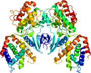 |
594キロバイト | 2 | |
| 2013年6月1日 (土) 12:49 | 3LL8.pdb (ファイル) |  |
792キロバイト | 1 | |
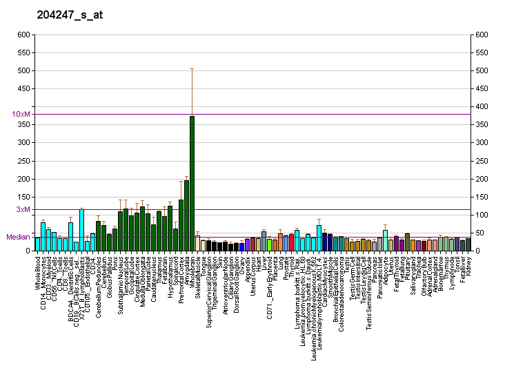
| 2013年6月1日 (土) 10:00 | PBB GE CDK5 204247 s at fs.png (ファイル) |  |
11キロバイト | {{Information |description={{en|Gene expression pattern of the CDK5 gene.}} |date={{Date|2007|11|3}} (original upload date) |source=Transferred from [http://en.wikipedia.org en.wikipedia]. Diagram created by [[:en:User... | 1 |
| 2013年6月1日 (土) 09:58 | PBB GE CDK5 204247 s at tn.png (ファイル) |  |
0バイト | {{Information |description={{en|Gene expression pattern of the CDK5 gene.}} |date={{Date|2007|11|3}} (original upload date) |source=Transferred from [http://en.wikipedia.org en.wikipedia]. Diagram created by [[:en:User... | 1 |
| 2013年5月19日 (日) 20:17 | Chair.cml (ファイル) | 2キロバイト | 1 | ||
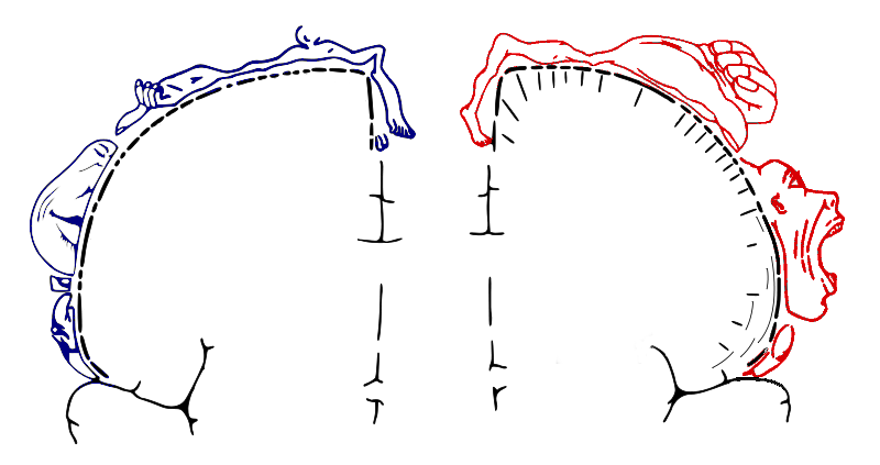
| 2013年5月16日 (木) 09:40 | Homunculus.png (ファイル) |  |
60キロバイト | {{Information |Description={{en|Homunculus: diagram showing position of regions of the human cortex corresponding to the respective afferent/efferent nerve region of the body. Blue: sensor cortex. Red: motor cortex.}} |Source=Wikimedia Commons (modified f | 1 |
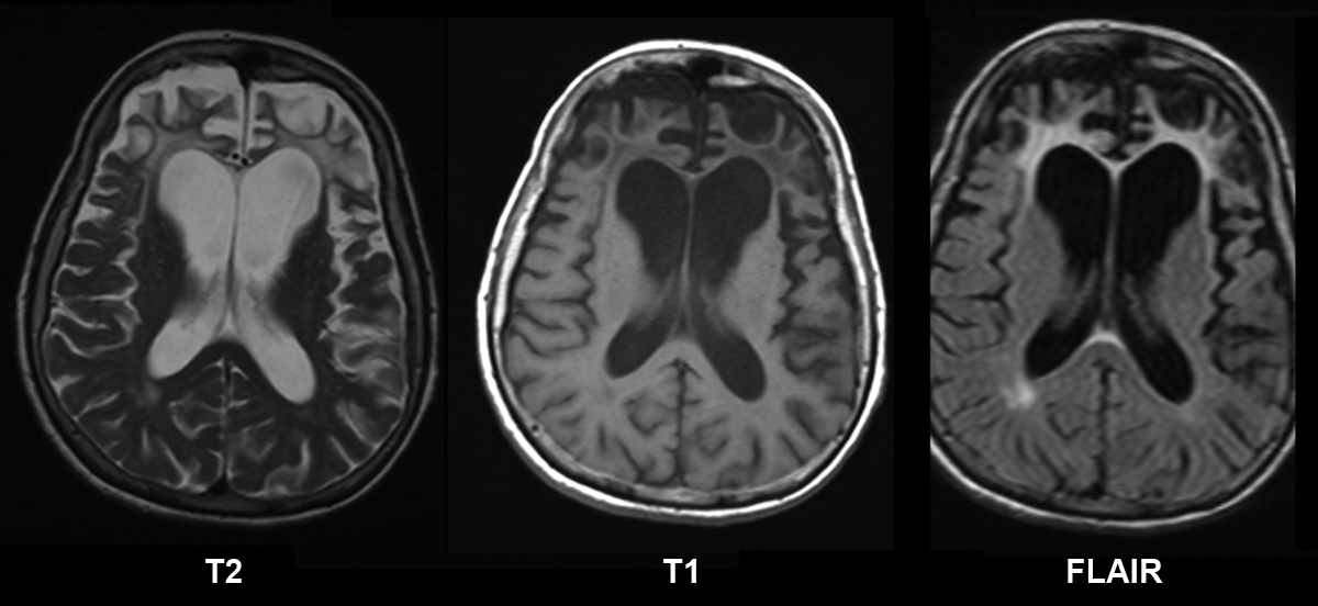
| 2013年5月12日 (日) 20:52 | Pick's disease.png (ファイル) |  |
223キロバイト | == {{int:filedesc}} == {{Information |Description={{en|1=Brain MRI of a female 65 y.o. white patient with Pick's disease. Cortex and white matter atrophy of the frontal lobes is clear visible. The MRI was done without contrast enhancement utilizing Magnet | 1 |
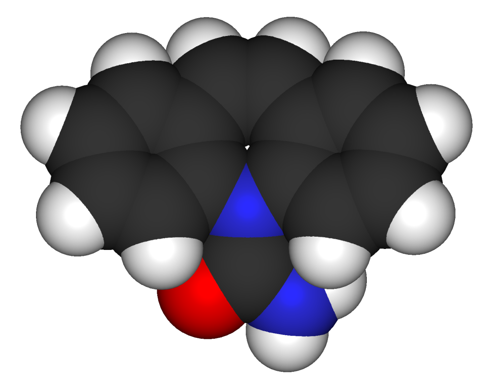
| 2013年5月5日 (日) 14:31 | Carbamazepine 3D.png (ファイル) |  |
136キロバイト | == {{int:filedesc}} == {{Information |Description= {{w|Space-filling model}} of {{w|carbamazepine}}. Created using [http://www.acdlabs.com/download/chemsk.html ACD/ChemSketch 10.0], [http://www.accelrys.com/products/downloads/ds_visualizer/index.html Acc | 1 |
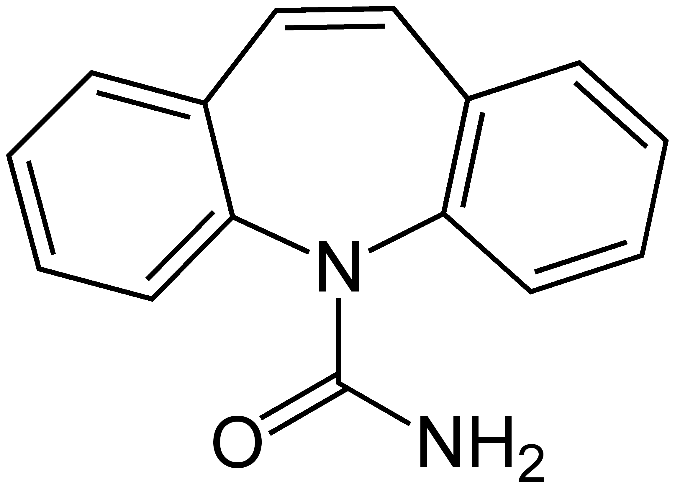
| 2013年5月5日 (日) 14:30 | Carbamazepine Structural Formulae.png (ファイル) |  |
30キロバイト | == {{int:filedesc}} == {{Information |Description={{en|1=Carbamazepine_Structural_Formulae}} |Source={{own}} |Author=Jü |Date=2009-11-22 |Permission= |other_versions= }} Category:Carbamazepine == {{int:license}} == {{PD-self}} | 1 |
| 2013年5月5日 (日) 14:28 | Sodium-valproate-2D-skeletal.png (ファイル) |  |
21キロバイト | {{SVG|chemical}} {{PD-self}} Category:Valproic acid Category:Sodium salts of organic compounds | 1 |
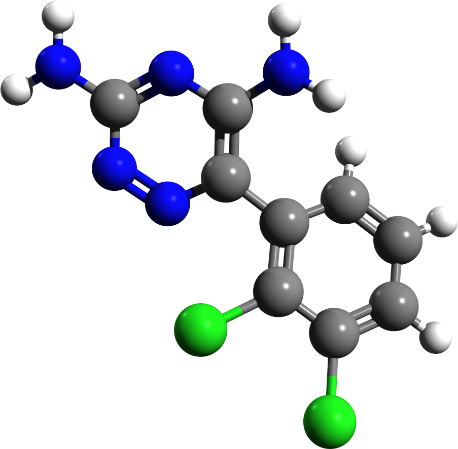
| 2013年5月5日 (日) 14:25 | Lamotrigine 3d structure.png (ファイル) |  |
152キロバイト | == {{int:filedesc}} == {{Information |Description={{en|1=Lamotrigine structure}} |Source={{own}} |Author=Giorgiogp2 |Date= |Permission= |other_versions= }} Category:Mood stabilizers == {{int:license-header}} == {{self|cc-by-sa-3.0 | 1 |
| 2013年5月5日 (日) 14:25 | Lamotrigine.svg (ファイル) |  |
16キロバイト | == {{int:filedesc}} == {{Information |Description={{en|1=2D structure of anticonvulsant drug lamotrigine}} |Source={{Own}} |Author=Harbin |Date=2009-01-04 |Permission={{PD-self}} |other_versions= }} Category:Anticonvulsants [[Category | 1 |
| 2013年5月5日 (日) 14:22 | Stylised Lithium Atom.svg (ファイル) |  |
10キロバイト | Made by Halfdan. SVG by Indolences Modification de Image:Stylised Lithium Atom.png par Liquid_2003. Represents Lithium-7. Black dots are electrons, red dots are protons and blue dots are neu | 1 |
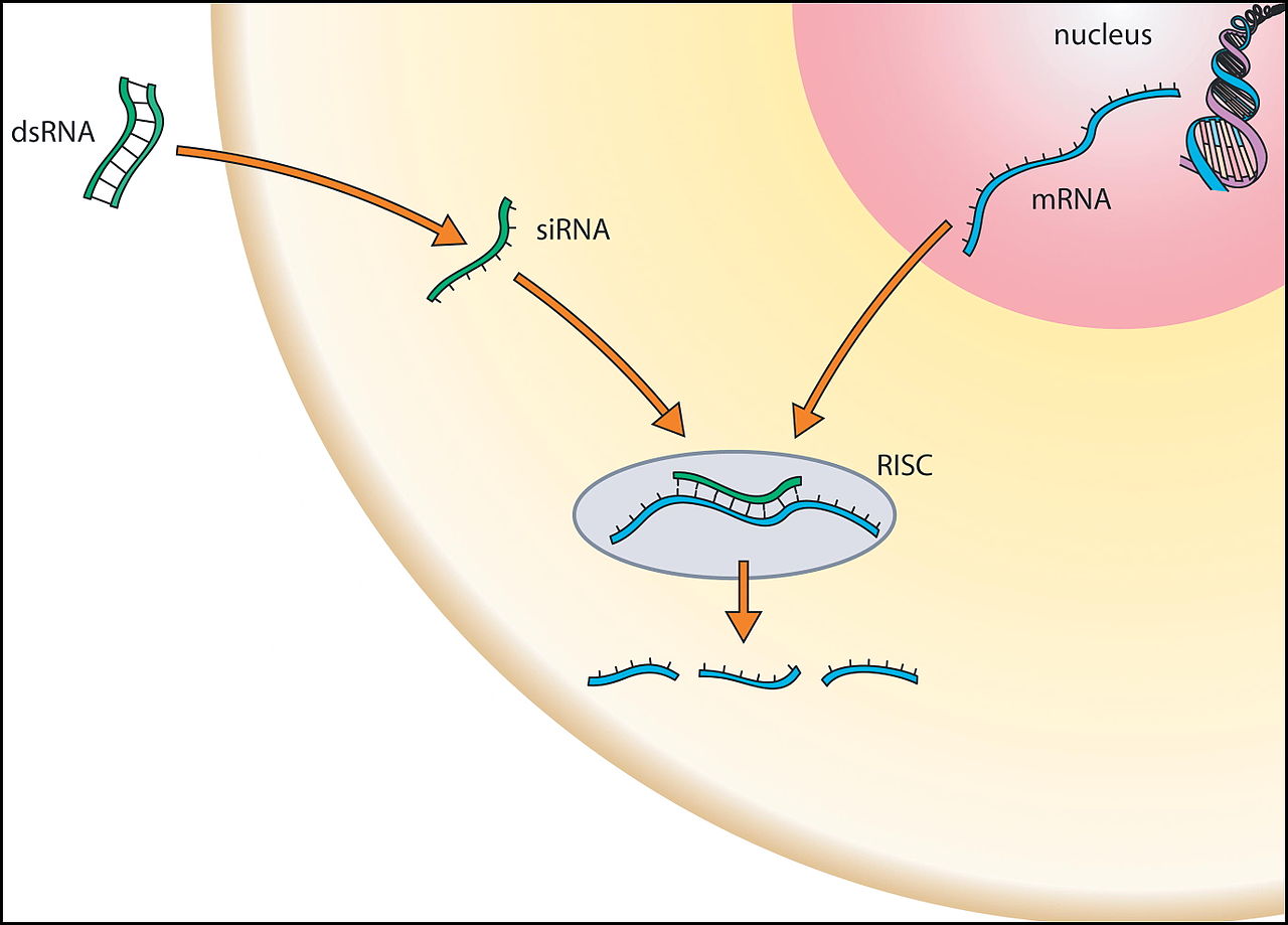
| 2013年5月3日 (金) 08:04 | RNA干渉.jpg (ファイル) |  |
79キロバイト | 2 | |
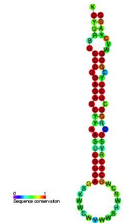
| 2013年5月3日 (金) 07:56 | RF00052.jpg (ファイル) |  |
26キロバイト | 2 | |
| 2013年4月13日 (土) 21:23 | Kakizawa morn repeat.png (ファイル) |  |
117キロバイト | 1 | |
| 2013年4月5日 (金) 23:26 | Anterior cingulate gyrus animation.gif (ファイル) |  |
1.63メガバイト | {{Information |Description={{en|1= Anterior cingulate gyrus. }} {{ja|1=前部帯状回。帯状回の前部。}} |Source=see below |Date=2011-01-04 |Author=see below |Permission={{Anatomography}} |other_versions=[[File:Anterior cingulate gyrus animation s | 1 |
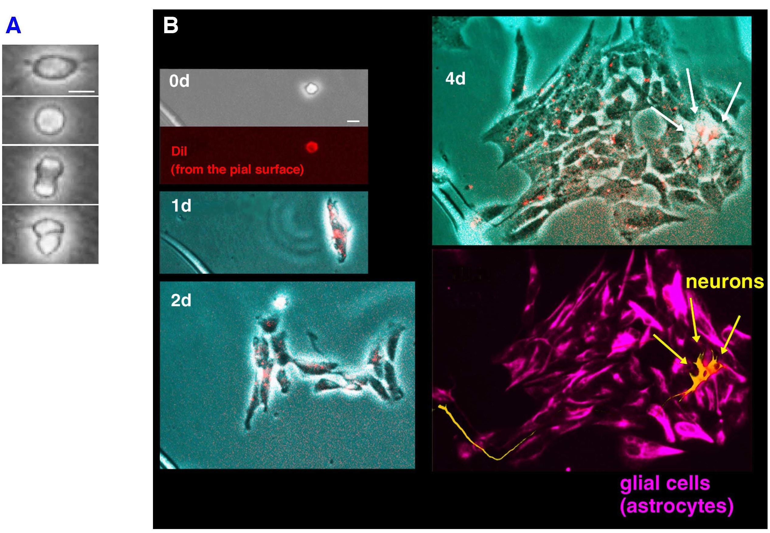
| 2013年3月26日 (火) 00:17 | タイムラプス解析データ例.jpg (ファイル) |  |
876キロバイト | 3 | |
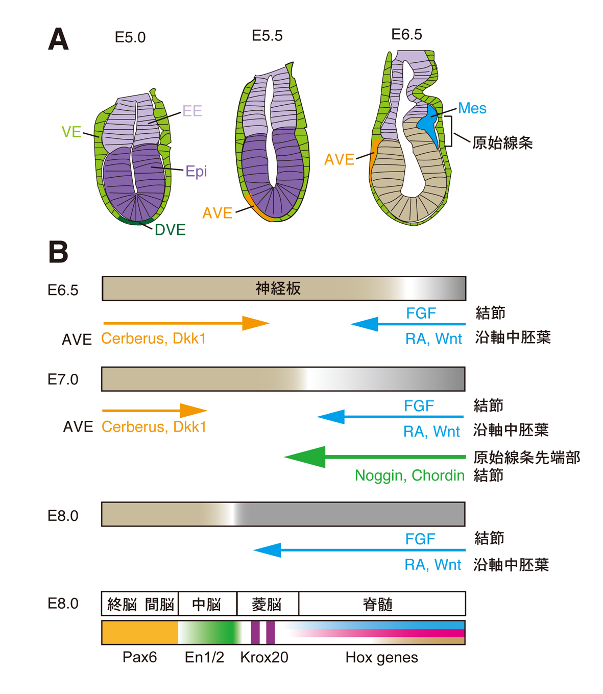
| 2013年3月23日 (土) 23:37 | 前後軸図1.jpg (ファイル) |  |
532キロバイト | 2 | |
| 2013年3月23日 (土) 23:35 | Masanoritakahashi fig2.jpg (ファイル) |  |
250キロバイト | 1 | |
| 2013年3月22日 (金) 20:56 | Higashijima fig2.png (ファイル) |  |
974キロバイト | 2 | |
| 2013年3月22日 (金) 20:56 | Higashijima fig1.png (ファイル) |  |
821キロバイト | 2 | |
| 2013年3月14日 (木) 10:18 | Protein CFTR PDB 1xmi.png (ファイル) |  |
607キロバイト | == {{int:filedesc}} == {{Information | Description={{en | 1=Structure of the CFTR protein. Based on PyMOL rendering of PDB [http://www.pdb.org/pdb/explore/explore.do?structureId=1xmi 1xmi].}} | Source={{own}} | Author=Emw | 1 |
| 2013年3月14日 (木) 10:01 | 1ots opm.gif (ファイル) |  |
130キロバイト | == {{int:filedesc}} == {{Information |Description=CIC voltage gated chloride channel from Escerischia coli. Protein image from OPM database {{cite journal |author=Dutzler R, Campbell EB, MacKinnon R |title=Gating the selectivity filter in ClC chloride | 1 |
| 2013年2月28日 (木) 21:18 | 1R02 crystallography.png (ファイル) |  |
143キロバイト | 1 | |
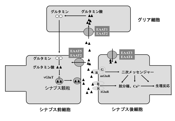
| 2013年2月24日 (日) 18:26 | Hayashi glutamate fig1.png (ファイル) |  |
175キロバイト | 4 | |
| 2013年2月24日 (日) 17:53 | Ritthausen 1861.pdf (ファイル) | 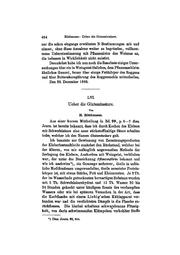 |
945キロバイト | 2 | |
| 2013年2月24日 (日) 12:59 | Hayashi glutamate fig2.png (ファイル) |  |
96キロバイト | 1 | |
| 2013年2月15日 (金) 20:30 | Corpus callosum.gif (ファイル) |  |
4.08メガバイト | {{Information |Description={{en|1=corpus callosum. Images are from Anatomography maintained by Life Science Databases(LSDB). }} {{ja|1=脳梁。 画像はLife Science Databases(LSDB)のAnatomographyというサイトより。}} |Source=from Anatomography | 1 |
| 2013年2月13日 (水) 21:28 | 3LL8.png (ファイル) |  |
229キロバイト | PDB entry 3ll8 Crystal structure of calcineurin in complex with akap79 peptide We found 1 Pubmed Central article that refers to 3ll8. You can see it here. Experiment type: Other, resolution 2.00Å Release date: 2011-01-12 Authors: Li, H., Hogan, P.G. | 1 |
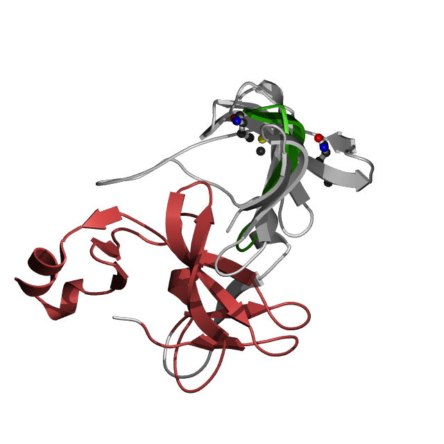
| 2013年2月12日 (火) 23:36 | 2p6b.png (ファイル) |  |
194キロバイト | From pfam database | 1 |
| 2013年2月5日 (火) 08:50 | MPTP Fig1.jpg (ファイル) |  |
40キロバイト | 2012年12月24日 (月)11:40の版へ差し戻し | 2 |
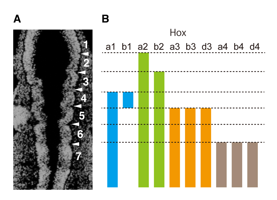
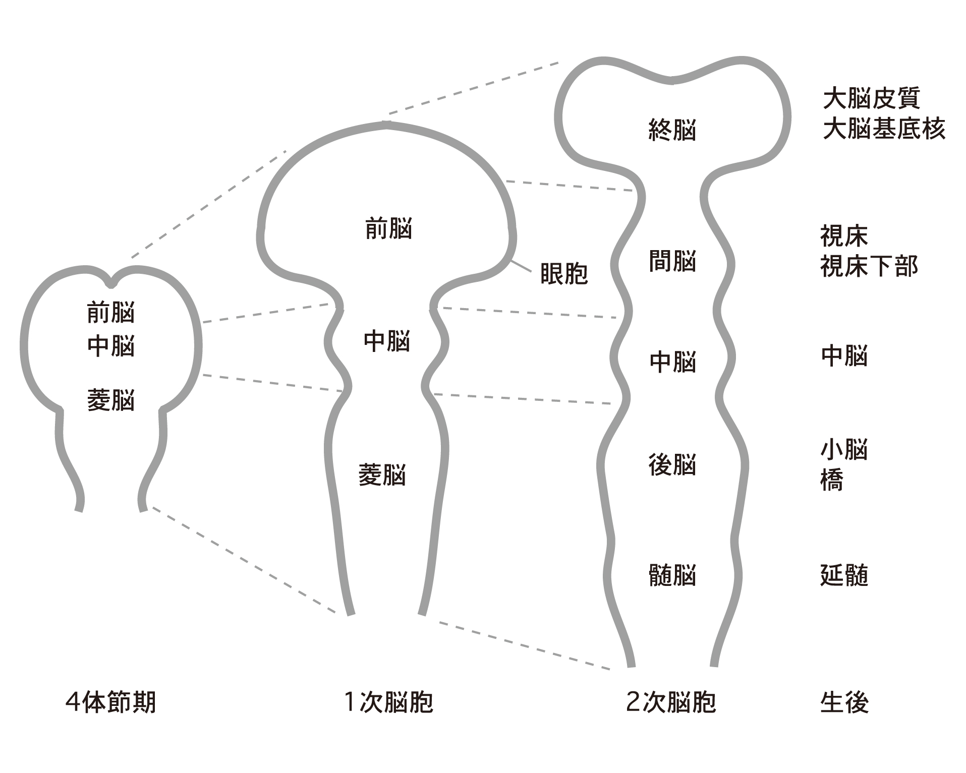
| 2013年2月3日 (日) 17:51 | 神経管図2.jpg (ファイル) |  |
465キロバイト | 2 | |
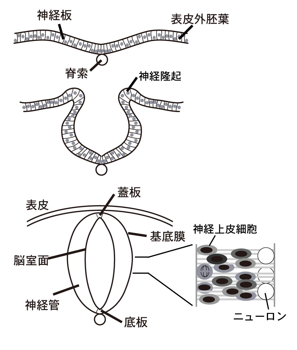
| 2013年2月3日 (日) 17:50 | 神経管図1.jpg (ファイル) |  |
370キロバイト | 2 | |
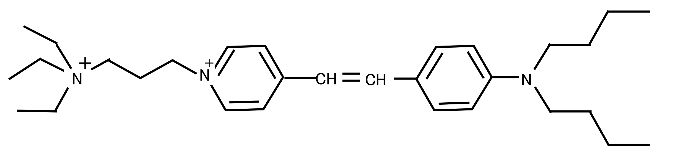
| 2013年2月1日 (金) 07:24 | FM1-43.png (ファイル) | 87キロバイト | 1 | ||
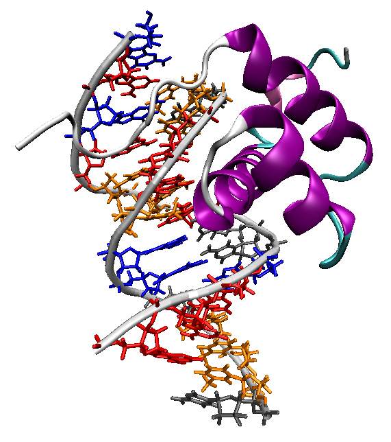
| 2013年1月24日 (木) 21:18 | Homeodomain-dna-1ahd.png (ファイル) |  |
179キロバイト | == {{int:filedesc}} == {{Information |Description=The ''Antennapedia'' homeodomain protein from ''Drosophila melanogaster'' bound to a DNA fragment, illustrating the binding interactions of the recognition helix and unstructured N-terminus with the DNA ma | 1 |
| 2013年1月18日 (金) 23:05 | Acetylcholine.svg (ファイル) |  |
6キロバイト | 1 | |
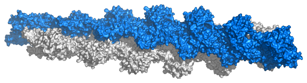
| 2013年1月17日 (木) 23:38 | Actin filament atomic model.png (ファイル) | 347キロバイト | 1 | ||
| 2013年1月15日 (火) 09:54 | Hayashi glutamate fig3.png (ファイル) |  |
58キロバイト | 1 | |
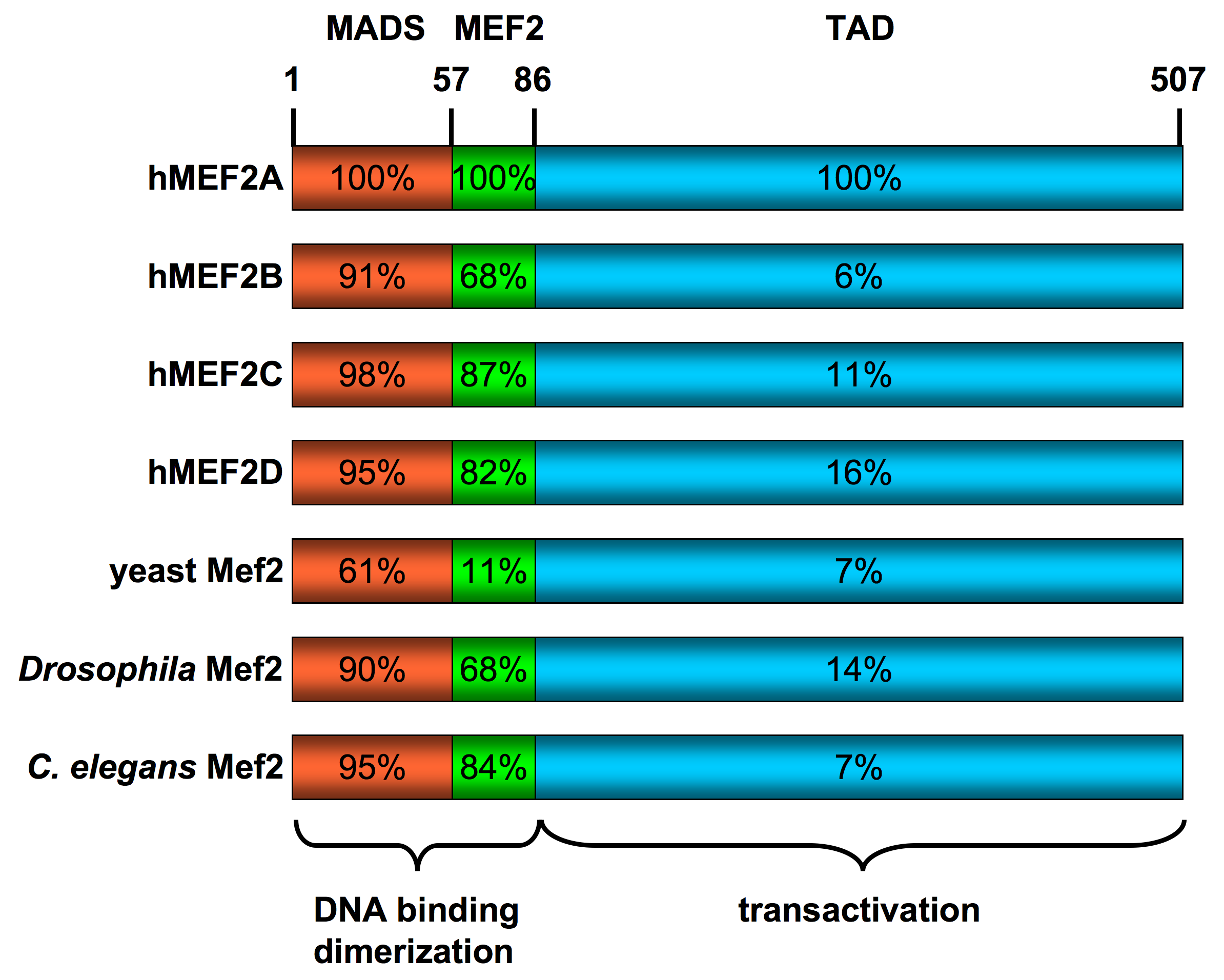
| 2013年1月13日 (日) 13:18 | MEF2 schematic.png (ファイル) |  |
159キロバイト | == Summary == {{Information |Description=Schematic diagram of the 1D sequence of the transcription factor MEF2 in various species. Numbering per cent homology all refer to the human MEF2A sequence. |Source=I created this work entirely by myself using Pow | 1 |
| 2013年1月13日 (日) 13:16 | Protein MEF2A PDB 1c7u.png (ファイル) |  |
303キロバイト | == {{int:filedesc}} == {{Information | Description={{en | 1=Structure of the MEF2A protein. Based on PyMOL rendering of PDB [http://www.pdb.org/pdb/explore/explore.do?structureId=1c7u 1c7u].}} | Source={{own}} | Author=Emw | 1 |
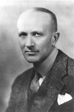
| 2012年12月8日 (土) 23:18 | Wilder Penfield.jpg (ファイル) |  |
17キロバイト | {{Information |Description={{en|Wilder Penfield Source: [http://www.archives.mcgill.ca/pictures/pr000632.GIF McGill University Archives] PORTRAIT: WILDER PENFIELD, DIRECTOR OF THE MONTREAL NEUROLOGICAL INSTITUTE, 1938-1960 Date: 1934CA Size: 003.5 X 0 | 1 |
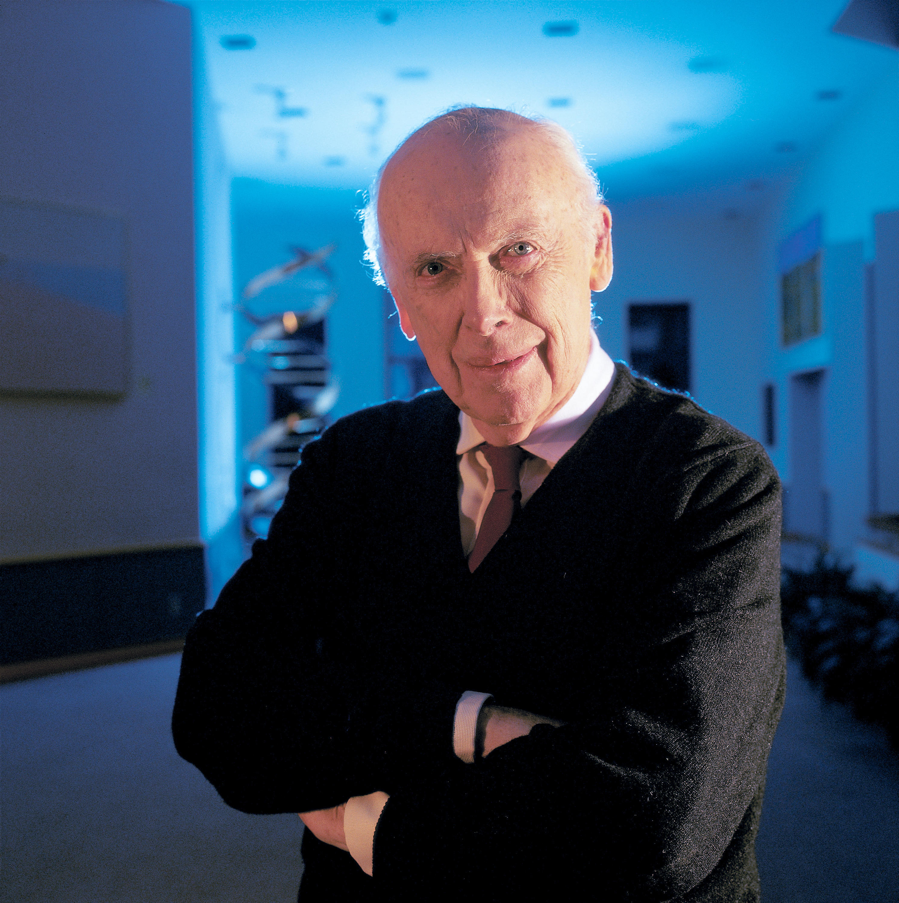
| 2012年12月8日 (土) 22:44 | James D Watson.jpg (ファイル) |  |
9.11メガバイト | == {{int:filedesc}} == {{Information |Description={{en|1=Nobel laureate Dr. James D. Watson, Chancellor, Cold Spring Harbor Laboratory. These images are freely available and may be used without special permission.}} |Source={{Derived from|James_D_Watson_G | 1 |
| 2012年12月8日 (土) 18:41 | Brodmann Cytoarchitectonics.PNG (ファイル) |  |
173キロバイト | == {{int:filedesc}} == {{Information |Description={{en|1=Cytoarchitectonics of human brain according to Brodmann (1909)}} |Source=Vergleichende Lokalisationslehre der Grosshirnrinde in ihren Prinzipien dargestellt auf Grund des Zellenbaues, Johann Ambrosi | 1 |
| 2012年12月6日 (木) 00:49 | Brain diagram without text.svg (ファイル) |  |
40キロバイト | == {{int:filedesc}} == {{Information |Description={{en|Principal lobes of the cerebrum viewed laterally. Figure 728 from Gray's Anatomy. 4 lines note sulci as follows *top center: Central sulcus *top right: Parieto-occipital sulcus *down left: Lateral sul | 1 |