腹側線条体
中村 加枝
関西医科大学 医学部
DOI:10.14931/bsd.7930 原稿受付日:2012年12月10日 原稿完成日:2012年12月13日
担当編集委員:渡辺 雅彦 (北海道大学大学院医学研究院 解剖学分野 解剖発生学教室)
英語名:ventral striatum 独:ventrales Striatum 仏:striatum ventral
腹側線条体はHeimer らによって提唱されたコンセプトで、側坐核とその周辺の領域を含む。入力元は腹内側前頭野・前頭眼窩野・島皮質といった皮質、扁桃体・海馬・視床髄板内核群を含む皮質下領域と中脳・脳幹のドーパミンをはじめとした神経伝達物質関連領域であり、出力は、大脳基底核の腹側淡蒼球と黒質網様部・緻密部、基底核外への投射としては外側視床下部・脚橋被蓋核・中心灰白質・分界条床核がある。これらの領域間の情報統合とドーパミンを中心とした神経伝達物質物質の作用により、快感・報酬・意欲・嗜癖・恐怖の情報処理に重要な役割を果たし、意思決定や薬物中毒の病態の責任部位であると考えられている。
定義と概要
腹側線条体はHeimerらによって提唱された概念で[1] 、側坐核(the nucleus accumbens NAcc)を中心として、それに接する前交連より吻側の尾状核腹内側から内包の腹側へ続く領域、被殻腹内側、外側嗅索に接する前有孔質(anterior perforated substance)を含む領域を含み、腹側は嗅結節(olfactory tubercle)に続く。
腹側線条体の大半を占める側坐核は薬物中毒・統合失調症・強迫性障害・注意欠陥多動性障害等の精神疾患との関連が指摘され、多くの知見が報告されている。

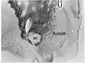
[2]より。
AcbSh, 側坐核shell; AcbC, 側坐核core; ac, 前交連
解剖
構築
腹側線条体は線条体の他の部分と多くの共通点を持ち、腹側線条体と背外側線条体(dorsolateral striatum)との境界については細胞構築・組織化学的には不明瞭であり、入力元によるところが大きい。腹側線条体・背側線条体どちらも皮質・視床・脳幹からの入力があるが、腹側線条体のみが扁桃体と海馬からの強い投射を受けている。
側坐核は解剖・機能的に背側線条体と共通点が多いcoreと、特異な点が多い三日月型のshellとから成る。組織化学的にはカルビンディン (calcium-binding protein, calbindin)染色でshellは薄くcoreは濃く染まる点が種を超えてみられる[2] 。ただし、coreと背側線条体の境界は不明瞭である。
Coreに比べ、shellはGluR1, GAP-43, アセチルコリンエステラーゼ、オピオイドμ受容体結合、セロトニン、サブスタンスPが豊富である。ドーパミントランスポーターはcoreも含め腹側線条体では背側線条体に比べて濃度が低い。これは、腹側線条体に主に投射するドーパミン細胞領域のdorsal tierでドーパミントランスポーターのmRNAが低値であることと一致する[3] 。
細胞形態学的には、背側線条体に比べ腹側線条体の細胞はやや小さく、密に分布する傾向にあり、striosome (patch)-matrix構造は背側線条体ほど明確に見られない[4][3] 。
入力
腹側線条体への入力元は皮質、皮質下入力、中脳ドーパミンがある(図2・3)。Shellはその他の腹側線条体にはない限定的な皮質入力を(32,24,14、Ia)受けており、特に、腹内側前頭野(ventromedial prefrontal cortex vmPFC)、島皮質Ia、扁桃体の諸核からoverlapした入力を受ける。Shellはその他の腹側線条体にはない出力があり、淡蒼球や黒質への投射に加えて視床下部や、分界条床核(the bed nucleus of the stria terminalis BNST)にも投射する[5] 。
皮質入力
霊長類では、shellには25・14・32野といった腹内側前頭野からの投射がある[6] 。これらに対し、腹側線条体の中心・外側部は前頭眼窩野(orbitofrontal cortex OFC、10・11・12・13野)からの投射を受ける。前頭眼窩野からの入力は嗅覚・味覚・内臓知覚など食べ物の感覚と関連し、これに視覚入力・後述の扁桃体からの入力も加わり、報酬情報に情動の要素が統合される。
前頭眼窩野とともに腹側線条体に感覚入力を送る皮質は島皮質である。島皮質は細胞構築学的・扱う知覚により
- Ia:顆粒細胞層を欠く無顆粒島(agranular insula嗅覚と自律神経反応)
- Id:亜顆粒島(dysgranular insula味覚と一部の視覚・体性感覚)
- Ig:全ての層構造が明瞭な顆粒島(granular insula体性感覚・聴覚と視覚)
に大別される。
腹側線条体はIaとIdから入力を受けるがIgからは受けない[7] 。Iaからの入力はshell内側と尾状核内側が最も強い。従って、shellで嗅覚と自律神経反応・腹内側前頭野からの入力が集約される。腹側線条体中心部へはIaおよびIdから投射する。したがって腹側線条体は島皮質と前頭眼窩野・腹内側前頭野から2重支配を受けている。
皮質下領域からの入力
扁桃体と海馬
扁桃体は辺縁系の一部で、外界の情動的な情報を伝達していると考えられている。この扁桃体から背側線条体への投射はほとんど存在しない。腹側線条体への投射は扁桃体の基底核と副基底核大細胞部からである.外側部からの腹側線条体への投射は少ない.
扁桃体は複数の核から成るが、嗅覚を除くすべてのmodalityの高度知覚処理に関わる基底外側複合体(basolateral nuclear group, BLNG, 外側核、基底核、副基底核 the basal and accessory basal nucleiから成る)がshellを除く腹側線条体への主な入力元である[8][9][10] 。Shellへの扁桃体からの入力はやや複雑であり、基底外側複合体に加え、皮質核・内側核・中心核からの入力がある。中心核には外界(BLNGを介して)や体内の状態(外側視床下部や脳幹を介して)の情報の入力があり、shellでは例えば外界の知覚入力と体内の例えば空腹という状態をあわせて、drive状態にすることに関与する。
海馬はさらに限られた領域であるshell へ投射し、扁桃体からの投射とoverlapしている[11][12] 。
視床
視床の内側部にある視床髄板内核群intralaminar thalamic nucleiは前頭葉内側・扁桃体・海馬に投射する辺縁系に属する視床核群であるが、腹側線条体はこれらの視床核からの投射を受ける[13] .Shellは束傍核parafascicular nucleusから投射を受ける。
腹側線条体はドーパミン細胞からの投射も強く受ける(下記回路の一部としての腹側線条体を参照。)
出力
腹側線条体からの出力投射先は,腹側淡蒼球(ventral pallidum VP)と黒質網様部・緻密部である[14] 。基底核外への投射としてはshellからの外側視床下部(the lateral hypothalamus)脚橋被蓋核(pedunculopontine nucleus)、中心灰白質、extended amygdalaの一部である分界条床核(BNST)がある。
回路の一部としての腹側線条体
腹側線条体はAlexanderらにより提唱された並列した複数の大脳皮質―線条体―淡蒼球・黒質―大脳皮質ループのうち、limbic loopの一部である[15] 。さらに、線条体―黒質―線条体「スパイラル」ループの一部としてもとらえられる。腹側線条体は複数の前頭葉皮質・扁桃体・海馬からの入力を集約して受け、さらに腹側被蓋野や黒質緻密部のドーパミン細胞に集約投射する。ドーパミン細胞はその後、線条体を中心に拡散的に投射を返す。このような線条体とドーパミン細胞の間の解剖・機能的な集約・拡散の繰り返しによって感覚入力に報酬情報が統合され、認知・行動の変化に影響を及ぼすメカニズムのひとつと考えられている[16] 。
機能
腹側線条体は種を超えて快感・報酬・意欲・嗜癖・恐怖の情報処理に重要な役割を果たし、報酬獲得行動や薬物中毒の病態の責任部位であると考えられている。近年は徐波睡眠との関連も明らかになってきている[17] 。
空間探索行動と行動選択
げっ歯類において、側坐核coreの障害実験により、この領域が空間情報・目標・教師信号などの入力を受けゴールにたどり着くための行動選択(空間探索行動, spatial navigation)に関わる[18][19] ことが示された。神経活動記録でも、側坐核coreのmedium spiny neuronが、いわゆるplace cellに相当する特定の居場所での発火を示すこと[20][21] 、さらにはその場所からの歩行の方向[22] によっても発火頻度が変化することが報告された。一方、shellの障害では、海馬からの入力が強いにもかかわらずそのような位置情報への影響は見られない。
情動や意欲、意思決定
ドーパミンやオピオイドの作用を変化させると、強迫的な選択行動等がひきおこされることから側坐核がhedonic(快楽的)な感情のプロセスに関与していることが示された。Berridgeらは、げっ歯類の側坐核の中で吻側―尾側軸の特定の異なる領域が、「快楽や報酬」領域と「恐怖や嫌悪」領域という異なる情動と対応していることを明らかにした[23]。
サルでもビククリンをごく少量注入して異なる腹側線条体領域の活動を局所的に障害すると、行動の抑制と意欲の低下・性的行動の亢進・異常な運動を繰り返す不安様行動が障害個所に特徴的に表れた[24] 。Shellの内側部は外側視床下部を抑制していて、これを障害すると摂食行動が引き起こされる[25] 。
線条体には、報酬と関連した感覚刺激や報酬そのものを予測的に期待する発火と、これらの後に反応する発火とが見られる。サルの電気生理学的実験では、背側線条体では課題の比較的前半つまり感覚刺激やその予測に関連した神経発火が見られるのに対し、腹側線条体では、課題の後半つまり報酬の予測や報酬を得た後に発火するものが多く観察されている[26][27] 。従って、腹側線条体は報酬を得るというゴールに達するため行動を起こす意欲driving forceの源となっている可能性がある。
ヒトの非侵襲的イメージングでは腹側線条体が報酬の予測・評価・報酬予測誤差の表現や、動機に基づいた学習に関与していることが示されたが、報酬の時間的予測については結論に差異がある[28][29][30][31][32] 。
Nicolaらは腹側被蓋野・内側前頭葉・扁桃体基底外側複合体の、側坐核単一細胞の発火への影響を調べた。ラットが音を弁別してレバー押しやnose pokeで反応することを学習すると、側坐核ニューロンは音に反応するが、腹側被蓋野・内側前頭葉・扁桃体からの入力をブロックすると側坐核ニューロンの発火が弱まり、弁別反応の正解率も低下する[33][34][35] 。したがって、これらの領域からの情報が側坐核で統合することが刺激―行動に必須であると結論付けられた。
一方、側坐核は報酬などの目的達成のためのオペラント条件付けそのものというより、現在進行中の行動から、一定の時間を経て別の行動に変化する過程に重要であるという意見がある[36][37][38] 。さらに、サルの腹側線条体細胞の特徴として、例えば視覚刺激→行動という単一試行の課題では課題に反応する細胞の割合は10%前後だが、複数のステップを経て報酬を得るような多試行報酬スケジュール課題では反応する細胞が60%前後と非常に多い。これらはスケジュールのうち特定の段階で、視覚手がかりへの応答や運動への応答報酬投与に応答する[39] 。
側坐核におけるドーパミンの作用については多くの知見がある。報酬を得られたらその行動を学習し、報酬を得られなかったら柔軟性を発揮して別の行動を選択する。この相反する行動決定の切り替えのメカニズムの少なくとも一部に、側坐核におけるphasicまたはtonicなドーパミンの作用のバランスが関与している。Phasicな作用は辺縁系(海馬)からの入力で主にドーパミンD1受容体を介する調節を受けている。Tonicな作用は前頭葉からの入力で主にドーパミンD2受容体を介する調節を受けている。腹側淡蒼球 (ventral pallidum, VP)は通常ドーパミン系をtonicに抑制している。海馬から興奮性の入力を受けると、側坐核は抑制性の投射をこのVPに送り、脱抑制機構によりドーパミン細胞をtonicに興奮させる。このtonicなドーパミン投射はD2受容体を介して前頭葉からの入力を抑制する。一方、phasicな作用は脚橋被蓋核からドーパミン細胞への入力による。これらの入力の側坐核でのバランスによって適切な行動の選択が可能となる[40] 。
関連項目
図2 腹側線条体への出入力。緑色は側坐核Core,青色はShellにより強く,白は同様の強度であることを示す[41] 。 A8, retrorubral area; ACC, anterior cingulate cortex; AId, dorsal anterior insular; AIv, ventral anterior insular; dHPC, dorsal hippocampus; dlVP, dorsolateral ventral pallidum; DRN, dorsal raphe nucleus; IL, infralimbic cortex; ILT, interlaminar nuclei of the thalamus; LC, locus coeruleus; LH, lateral hypothalamus; LPO, lateral preoptic area; NTS, nucleus of the solitary tract; PL, prelimbic cortex; PPN, pedunculopontine nucleus; PVT, paraventricular nucleus of the thalamus; vlVP, ventrolateral ventral pallidum; vmVP, ventromedial ventral pallidum; SNc, substantia nigra pars compacta; SNpr, substantia nigra pars reticulata.
図3 霊長類における腹側線条体への出入力[42] より改変 赤矢印: vmPFCからの入力; 濃オレンジ矢印: OFCからの入力; 淡オレンジ矢印: dACCからの入力;黄色矢印: dPFCからの入力;茶色矢印:その他 Amy,amygdala; dACC,dorsal anterior cingulate cortex; dPFC, dorsal prefrontal cortex; Hipp,hippocampus; LHb,lateral habenula; hypo,hypothalamus; OFC,orbital frontal cortex; PPT,pedunculopontine nucleus; S,shell, SNc¼substantia nigra, pars compacta; STN¼subthalamic nucleus.; Thal¼thalamus; VP,ventral pallidum; VTA,ventral tegmental area; vmPFC,ventral medial prefrontal cortex.Ia,agranular insula;Id,dysgranular insula;Ig,granular insula
1.定義と概要 腹側線条体(ventral striatum 以下VS) は側坐核(the nucleus accumbens NAc)を中心として、それに接する前交連より吻側の尾状核腹内側から内包の腹側へ続く領域、被殻腹内側、外側嗅索に接する前有孔質(anterior perforated substance)を含む領域を含み、腹側は嗅結節(olfactory tubercle)に続く。VSの大半を占める側坐核は薬物中毒・統合失調症・強迫性障害・注意欠陥多動性障害等の精神疾患との関連が指摘され、多くの知見が報告されている。
VSは線条体の他の部分と多くの共通点を持ち、VSと背外側線条体(dorsolateral striatum 以下DS)との境界については細胞構築・組織化学的には不明瞭であり、入力元によるところが大きい。VS・DSどちらも皮質・視床・脳幹からの入力があるが、VSのみが扁桃体と海馬からの強い投射を受けている。 側坐核は解剖・機能的にDSと共通点が多いcoreと、特異な点が多い三日月型のshellとから成る。組織化学的にはcalbindin (calcium-binding protein)染色でshellは薄くcoreは濃く染まる点が種を超えてみられる[2] 。ただし、coreとDSの境界は不明瞭である。coreに比べ、shellはGluRl, GAP-43, acetylcholinesterase,μ receptor binding, serotonin, substance Pが豊富である。Dopamine transporterはcoreも含めVSではDSに比べて濃度が低い。これは、VSに主に投射するドーパミン細胞領域のdorsal tierでdopamine transporterの mRNAが低値であることと一致する[3] 。細胞形態学的には、DSに比べVSの細胞はやや小さく、密に分布する傾向にあり、striosome (patch)-matrix構造はDSほど明確に見られない[4] ;[3]
2.入力元 (図2・3) 概要:VSへの入力元は皮質、皮質下入力、背側中脳ドーパミン細胞領域がある。Shellはその他のVSにはない限定的な皮質特に、内側・腹内側前頭野(32,24,25、14野),島皮質Iaからの入力を受けている。これが扁桃体の諸核からの入力とoverlapする。また、中脳ドパミン細胞のdorsal tier部からの入力を受ける。Shellはその他のVSにはない出力があり、淡蒼球や黒質への投射に加えて視床下部や・分界条床核(the bed nucleus of the stria terminalis BNST)にも投射する[5] 。 1)皮質入力 霊長類では、shell部分は32,24,25, 14野といった内側・腹内側前頭野と島皮質からの投射がある[6] 。shell内側部は特に25,14、Iaからの投射が強い。これらに対し、VSの中心・外側部は前頭眼窩野(orbitofrontal cortex OFC、10・11・12・13野)からの投射を受ける。OFCからの入力は嗅覚・味覚・内臓知覚など食べ物の感覚と関連し、これに視覚入力・後述の扁桃体からの入力も加わり、報酬情報に情動の要素が統合される。 OFCとともにVSに感覚入力を送る皮質は島皮質(Insula)である。島皮質は細胞構築学的・扱う知覚により(Ia)顆粒細胞層を欠く無顆粒島(agranular insula嗅覚と自律神経反応)、(Id)亜顆粒島(dysgranular insula味覚と一部の視覚・体性感覚)、(Ig)全ての層構造が明瞭な顆粒島(granular insula体性感覚・聴覚と視覚)に大別される。VSはIaとIdから入力を受けるがIgからは受けない[7] 。Iaからの入力はshell内側と尾状核内側が最も強い。従って、shellで嗅覚と自律神経反応・vmPFCからの入力が集約される。VS中心部へはIaおよびIdから投射する。したがってVS は島皮質とOFC・vmPFCから2重支配を受けている。
2)皮質下領域の入力 扁桃体と海馬:扁桃体は辺縁系の一部で、外界の情動的な情報を伝達していると考えられている。この扁桃体からDSへの直接的な投射はほとんど存在しない。VSへの投射は扁桃体の基底核と副基底核大細胞部からである。外側部からのVSへの投射は少ない. 扁桃体は複数の核から成るが、嗅覚を除くすべてのmodalityの高度知覚処理に関わる基底外側複合体(basolateral nuclear group, BLNG, 外側核、基底核、副基底核the basal and accessory basal nucleiから成る)がshellを除くVSへの主な入力元である[8][9][10] 。Shellへの扁桃体からの入力はやや複雑であり、基底外側複合体に加え、皮質核・内側核・中心核からの入力がある。中心核には外界(BLNGを介して)や体内の状態(外側視床下部や脳幹を介して)の情報の入力があり、shellでは例えば外界の知覚入力と体内の例えば空腹という状態をあわせて、drive状態にすることに関与する。 海馬はさらに限られた領域であるshell へ投射し、扁桃体からの投射とoverlapしている[11][12] 。
視床: VSは正中視床核群midline nuclei(室傍核paraventricular nucleus、結合核reuniens nucleus、菱形核 rhomboid nucleus、紐傍核paratenial nucleus)と視床髄板内核群intralaminar thalamic nucleiに属する束傍核parafascicular nucleus内側部からの投射を受ける。正中視床核群は前頭葉内側・扁桃体・海馬に投射する辺縁系に属する視床核群である (Gimenez-Amaya et al., 1995)。
中脳ドーパミン細胞群: 中脳ドーパミン細胞は、背側黒質緻密部・腹側被蓋野からなり、calbindin positive なdorsal tierと、 densocellular cell group・黒質網様体に入り込むcolumn部からなりcalbindin negativeなventral tier がある。DSはventral tierから投射を受け、dorsal tierからは投射を受けない。これに対し、VSはdorsal tierおよびventral tier のdensocellular cell groupから投射を受け、column部からは受けない[43] 。
3.出力先 VSからの投射先は,腹側淡蒼球(ventral pallidum VP)と黒質網様部・緻密部である[14] 。さらに、shellからは外側視床下部(the lateral hypothalamus)脚橋被蓋核(pedunculopontine nucleus)・中心灰白質・extended amygdalaの一部である分界条床核(BNST)への投射がある。
4.回路の一部としての腹側線条体 VSはAlexanderらにより提唱された並列した複数の大脳皮質―線条体―淡蒼球・黒質―大脳皮質ループのうち、limbic loopの一部である[15] 。さらに、線条体―黒質―線条体「スパイラルループ」の一部としてもとらえられる。VSは複数の前頭葉皮質・扁桃体・海馬からの入力を集約して受け、さらに腹側被蓋野や黒質緻密部のドーパミン細胞に集約投射する。ドーパミン細胞はその後、線条体を中心に拡散的に投射を返す。このような線条体とドーパミン細胞の間の解剖・機能的な集約・拡散の繰り返しによって感覚入力に報酬情報が統合され、認知・行動の変化に影響を及ぼすメカニズムのひとつと考えられている[16] 。
5.機能 腹側線条体は種を超えて、行動選択に加え、快感・報酬・意欲・嗜癖・恐怖の情報処理に重要な役割を果たし、報酬獲得行動や薬物中毒の病態の責任部位であると考えられている。近年は徐波睡眠との関連も明らかになってきている[17] 。
1) spatial navigationと行動選択 げっ歯類において、側坐核coreの障害実験により、この領域が空間情報・目標・教師信号などの入力を受けゴールにたどり着くための行動選択(spatial navigation)に関わることが示された[18][19] 。神経活動記録でも、側坐核coreのmedium spiny neuronが、いわゆるplace cellに相当する特定の居場所での発火を示すこと[20] 、さらにはその場所からの歩行の方向[22] によっても発火頻度が変化することが報告された。一方、shellの障害では、海馬からの入力が強いにもかかわらずそのような位置情報への影響は見られない。
2) 情動・意欲・意思決定 ドーパミンやオピオイドの作用により強迫的な選択行動等がひきおこされることから、側坐核がhedonic(快楽的)な感情のプロセスに関与していることが示された。Berridgeらは、げっ歯類の側坐核の中で吻側―尾側軸の特定の異なる領域が、「快楽や報酬」領域と「恐怖や嫌悪」領域という異なる情動と対応していると報告した[23] 。サルでもbicucullineをごく少量注入して異なるVS領域の活動を局所的に変化させると、行動の抑制と意欲の低下・性的行動の亢進・異常な運動を繰り返す不安様行動が注入個所に特異的に表れた[24] 。Shellの内側部は外側視床下部を抑制していて、これを障害すると摂食行動が引き起こされる[25] 。
線条体には、報酬と関連した感覚刺激や報酬そのものを予測的に期待する発火と、これらの後に反応する発火とが見られる。サルの電気生理学的実験では、DSでは課題の比較的前半つまり感覚刺激やその予測に関連した神経発火が見られるのに対し、VSでは、課題の後半つまり報酬の予測や報酬を得た後に発火するものが多く観察されている[26][27] 。従って、VSは報酬を得るというゴールに達するため行動を起こす意欲driving forceの源となっている可能性がある。 ヒトの非侵襲的イメージングではVSが報酬の予測・評価・報酬予測誤差の表現や、動機に基づいた学習に関与していることが示された。報酬の時間的予測については結論に差異がある[28][29][30][44] 。
Nicolaらは腹側被蓋野・内側前頭葉・扁桃体基底外側複合体の、側坐核単一細胞の発火への影響を調べた。ラットが音を弁別してレバー押しやnose pokeで反応することを学習すると、側坐核ニューロンは音に反応するが、腹側被蓋野・内側前頭葉・扁桃体からの入力をブロックすると側坐核ニューロンの発火は弱まり、弁別反応の正解率も低下する[33] [34] ; [35] 。したがって、これらの領域からの情報が側坐核で統合することが刺激―行動に必須であると結論付けられた。 一方、側坐核は報酬などの目的達成のためのオペラント条件付けそのものというより、現在進行中の行動から、一定の時間を経て別の行動に変化する過程に重要であるという意見がある[36][37][38] 。さらに、サルのVS細胞の特徴として、例えば視覚刺激―>行動という単一試行の課題では課題に反応する細胞の割合は10%前後だが、複数のステップを経て報酬を得るような多試行報酬スケジュール課題では反応する細胞が60%前後と非常に多い。これらはスケジュールのうち特定の段階で、視覚手がかりへの応答や運動への応答報酬投与に応答する[39] 。
側坐核におけるドーパミンの作用については多くの知見がある。報酬を得られたらその行動を学習し、報酬を得られなかったら柔軟性を発揮して別の行動を選択する。この相反する行動決定の切り替えのメカニズムの少なくとも一部に、側坐核におけるphasicまたはtonicなドーパミンの作用のバランスが関与している。Phasicな作用は辺縁系(海馬)からの入力で主にドーパミンD1受容体を介する調節を受けている。Tonicな作用は前頭葉からの入力で主にドーパミンD2受容体を介する調節を受けている。VP(ventral pallidum)は通常ドーパミン系をtonicに抑制している。海馬から興奮性の入力を受けると、側坐核は抑制性の投射をこのVPに送り、脱抑制機構によりドーパミン細胞をtonicに興奮させる。このtonicなドーパミン投射はD2受容体を介して前頭葉からの入力を抑制する。一方、phasicな作用は脚橋被蓋核からドーパミン細胞への入力による。これらの入力の側坐核でのバランスによって適切な行動の選択が可能となる[40] 。
図1 Calbindin染色によるげっ歯類の腹側線条体の区分[2] 。
AcbSh,Accumbens shell; AcbC,AcbSh core; ac,anterior commissure
図2 げっ歯類における腹側線条体への出入力。緑色は側坐核core,青色はshellにより強く,白は同様の強度であることを示す。[41] より改変) A8, retrorubral area; ACC, anterior cingulate cortex; AId, dorsal anterior insular; AIv, ventral anterior insular; dHPC, dorsal hippocampus; dlVP, dorsolateral ventral pallidum; DRN, dorsal raphe nucleus; IL, infralimbic cortex; ILT, interlaminar nuclei of the thalamus; LC, locus coeruleus; LH, lateral hypothalamus; LPO, lateral preoptic area; NTS, nucleus of the solitary tract; PL, prelimbic cortex; PPN, pedunculopontine nucleus; PVT, paraventricular nucleus of the thalamus; vlVP, ventrolateral ventral pallidum; vmVP, ventromedial ventral pallidum; SNc, substantia nigra pars compacta; SNpr, substantia nigra pars reticulata.
図3 霊長類における腹側線条体への出入力。[42] より改変) 赤矢印: vmPFCからの入力; 濃オレンジ矢印: OFCからの入力; 淡オレンジ矢印: dACCからの入力;淡黄色矢印:dPFCからの入力;茶色矢印:その他 Amy,amygdala; dACC,dorsal anterior cingulate cortex; dPFC, dorsal prefrontal cortex; Hipp,hippocampus; LHb,lateral habenula; hypo,hypothalamus; OFC,orbital frontal cortex; PPT,pedunculopontine nucleus; S,shell, SNc,substantia nigra, pars compacta; STN,subthalamic nucleus; Thal,thalamus; VP,ventral pallidum; VTA,ventral tegmental area; vmPFC,ventral medial prefrontal cortex.Ia,agranular insula;Id,dysgranular insula;Ig,granular insula
- ↑ Heimer, L.
The subcortical projections of the allocortex: similarities in the neural associations of the hippocampus, the piriform cortex, and the neocortex.
In Golgi Centennial Symposium: Perspectives in Neurobiology M Santini, Ed: 177-193 Raven Press New York. 1975 - ↑ 2.0 2.1 2.2 2.3
Groenewegen, H.J., Wright, C.I., Beijer, A.V., & Voorn, P. (1999).
Convergence and segregation of ventral striatal inputs and outputs. Annals of the New York Academy of Sciences, 877, 49-63. [PubMed:10415642] [WorldCat] [DOI] - ↑ 3.0 3.1 3.2 3.3
Haber, S.N., & McFarland, N.R. (1999).
The concept of the ventral striatum in nonhuman primates. Annals of the New York Academy of Sciences, 877, 33-48. [PubMed:10415641] [WorldCat] [DOI] - ↑ 4.0 4.1
Holt, D.J., Graybiel, A.M., & Saper, C.B. (1997).
Neurochemical architecture of the human striatum. The Journal of comparative neurology, 384(1), 1-25. [PubMed:9214537] [WorldCat] [DOI] 引用エラー: 無効な<ref>タグ; name "Holt1997"が異なる内容で複数回定義されています - ↑ 5.0 5.1
Humphries, M.D., & Prescott, T.J. (2010).
The ventral basal ganglia, a selection mechanism at the crossroads of space, strategy, and reward. Progress in neurobiology, 90(4), 385-417. [PubMed:19941931] [WorldCat] [DOI] - ↑ 6.0 6.1
Carmichael, S.T., & Price, J.L. (1995).
Limbic connections of the orbital and medial prefrontal cortex in macaque monkeys. The Journal of comparative neurology, 363(4), 615-641. [PubMed:8847421] [WorldCat] [DOI] - ↑ 7.0 7.1
Chikama, M., McFarland, N.R., Amaral, D.G., & Haber, S.N. (1997).
Insular cortical projections to functional regions of the striatum correlate with cortical cytoarchitectonic organization in the primate. The Journal of neuroscience : the official journal of the Society for Neuroscience, 17(24), 9686-705. [PubMed:9391023] [WorldCat] - ↑ 8.0 8.1
Iwai, E., & Yukie, M. (1987).
Amygdalofugal and amygdalopetal connections with modality-specific visual cortical areas in macaques (Macaca fuscata, M. mulatta, and M. fascicularis). The Journal of comparative neurology, 261(3), 362-87. [PubMed:3611417] [WorldCat] [DOI] - ↑ 9.0 9.1
Pitkänen, A., Savander, V., & LeDoux, J.E. (1997).
Organization of intra-amygdaloid circuitries in the rat: an emerging framework for understanding functions of the amygdala. Trends in neurosciences, 20(11), 517-23. [PubMed:9364666] [WorldCat] [DOI] - ↑ 10.0 10.1
Russchen, F.T., Amaral, D.G., & Price, J.L. (1987).
The afferent input to the magnocellular division of the mediodorsal thalamic nucleus in the monkey, Macaca fascicularis. The Journal of comparative neurology, 256(2), 175-210. [PubMed:3549796] [WorldCat] [DOI] 引用エラー: 無効な<ref>タグ; name "Russchen1987"が異なる内容で複数回定義されています - ↑ 11.0 11.1
Friedman, D.P., Aggleton, J.P., & Saunders, R.C. (2002).
Comparison of hippocampal, amygdala, and perirhinal projections to the nucleus accumbens: combined anterograde and retrograde tracing study in the Macaque brain. The Journal of comparative neurology, 450(4), 345-65. [PubMed:12209848] [WorldCat] [DOI] - ↑ 12.0 12.1
Saunders, R.C., & Rosene, D.L. (1988).
A comparison of the efferents of the amygdala and the hippocampal formation in the rhesus monkey: I. Convergence in the entorhinal, prorhinal, and perirhinal cortices. The Journal of comparative neurology, 271(2), 153-84. [PubMed:2454246] [WorldCat] [DOI] - ↑
Giménez-Amaya, J.M., McFarland, N.R., de las Heras, S., & Haber, S.N. (1995).
Organization of thalamic projections to the ventral striatum in the primate. The Journal of comparative neurology, 354(1), 127-49. [PubMed:7542290] [WorldCat] [DOI] - ↑ 14.0 14.1
Haber, S.N., Wolfe, D.P., & Groenewegen, H.J. (1990).
The relationship between ventral striatal efferent fibers and the distribution of peptide-positive woolly fibers in the forebrain of the rhesus monkey. Neuroscience, 39(2), 323-38. [PubMed:1708114] [WorldCat] [DOI] - ↑ 15.0 15.1 Resource not found in PubMed.
- ↑ 16.0 16.1
Haber, S.N., Fudge, J.L., & McFarland, N.R. (2000).
Striatonigrostriatal pathways in primates form an ascending spiral from the shell to the dorsolateral striatum. The Journal of neuroscience : the official journal of the Society for Neuroscience, 20(6), 2369-82. [PubMed:10704511] [PMC] [WorldCat] - ↑ 17.0 17.1
Oishi, Y., Xu, Q., Wang, L., Zhang, B.J., Takahashi, K., Takata, Y., ..., & Lazarus, M. (2017).
Slow-wave sleep is controlled by a subset of nucleus accumbens core neurons in mice. Nature communications, 8(1), 734. [PubMed:28963505] [PMC] [WorldCat] [DOI] - ↑ 18.0 18.1
Sargolini, F., Florian, C., Oliverio, A., Mele, A., & Roullet, P. (2003).
Differential involvement of NMDA and AMPA receptors within the nucleus accumbens in consolidation of information necessary for place navigation and guidance strategy of mice. Learning & memory (Cold Spring Harbor, N.Y.), 10(4), 285-92. [PubMed:12888547] [PMC] [WorldCat] [DOI] - ↑ 19.0 19.1
Ploeger, G.E., Spruijt, B.M., & Cools, A.R. (1994).
Spatial localization in the Morris water maze in rats: acquisition is affected by intra-accumbens injections of the dopaminergic antagonist haloperidol. Behavioral neuroscience, 108(5), 927-34. [PubMed:7826515] [WorldCat] [DOI] - ↑ 20.0 20.1
Shibata, R., Mulder, A.B., Trullier, O., & Wiener, S.I. (2001).
Position sensitivity in phasically discharging nucleus accumbens neurons of rats alternating between tasks requiring complementary types of spatial cues. Neuroscience, 108(3), 391-411. [PubMed:11738254] [WorldCat] [DOI] 引用エラー: 無効な<ref>タグ; name "Shibata2001"が異なる内容で複数回定義されています - ↑
Mulder, A.B., Tabuchi, E., & Wiener, S.I. (2004).
Neurons in hippocampal afferent zones of rat striatum parse routes into multi-pace segments during maze navigation. The European journal of neuroscience, 19(7), 1923-32. [PubMed:15078566] [WorldCat] [DOI] - ↑ 22.0 22.1
Peoples, L.L., Gee, F., Bibi, R., & West, M.O. (1998).
Phasic firing time locked to cocaine self-infusion and locomotion: dissociable firing patterns of single nucleus accumbens neurons in the rat. The Journal of neuroscience : the official journal of the Society for Neuroscience, 18(18), 7588-98. [PubMed:9736676] [PMC] [WorldCat] - ↑ 23.0 23.1
Berridge, K.C., & Kringelbach, M.L. (2008).
Affective neuroscience of pleasure: reward in humans and animals. Psychopharmacology, 199(3), 457-80. [PubMed:18311558] [PMC] [WorldCat] [DOI] - ↑ 24.0 24.1
Worbe, Y., Baup, N., Grabli, D., Chaigneau, M., Mounayar, S., McCairn, K., ..., & Tremblay, L. (2009).
Behavioral and movement disorders induced by local inhibitory dysfunction in primate striatum. Cerebral cortex (New York, N.Y. : 1991), 19(8), 1844-56. [PubMed:19068490] [WorldCat] [DOI] - ↑ 25.0 25.1
Kelley, A.E., Baldo, B.A., Pratt, W.E., & Will, M.J. (2005).
Corticostriatal-hypothalamic circuitry and food motivation: integration of energy, action and reward. Physiology & behavior, 86(5), 773-95. [PubMed:16289609] [WorldCat] [DOI] - ↑ 26.0 26.1 Resource not found in PubMed.
- ↑ 27.0 27.1
Nakamura, K., Santos, G.S., Matsuzaki, R., & Nakahara, H. (2012).
Differential reward coding in the subdivisions of the primate caudate during an oculomotor task. The Journal of neuroscience : the official journal of the Society for Neuroscience, 32(45), 15963-82. [PubMed:23136434] [PMC] [WorldCat] [DOI] - ↑ 28.0 28.1
Knutson, B., Adams, C.M., Fong, G.W., & Hommer, D. (2001).
Anticipation of increasing monetary reward selectively recruits nucleus accumbens. The Journal of neuroscience : the official journal of the Society for Neuroscience, 21(16), RC159. [PubMed:11459880] [PMC] [WorldCat] 引用エラー: 無効な<ref>タグ; name "Knutson2001"が異なる内容で複数回定義されています - ↑ 29.0 29.1
Pagnoni, G., Zink, C.F., Montague, P.R., & Berns, G.S. (2002).
Activity in human ventral striatum locked to errors of reward prediction. Nature neuroscience, 5(2), 97-8. [PubMed:11802175] [WorldCat] [DOI] - ↑ 30.0 30.1
Tanaka, S.C., Doya, K., Okada, G., Ueda, K., Okamoto, Y., & Yamawaki, S. (2004).
Prediction of immediate and future rewards differentially recruits cortico-basal ganglia loops. Nature neuroscience, 7(8), 887-93. [PubMed:15235607] [WorldCat] [DOI] 引用エラー: 無効な<ref>タグ; name "Tanaka2004"が異なる内容で複数回定義されています - ↑
O'Doherty, J., Dayan, P., Schultz, J., Deichmann, R., Friston, K., & Dolan, R.J. (2004).
Dissociable roles of ventral and dorsal striatum in instrumental conditioning. Science (New York, N.Y.), 304(5669), 452-4. [PubMed:15087550] [WorldCat] [DOI] - ↑
Kuhnen, C.M., & Knutson, B. (2005).
The neural basis of financial risk taking. Neuron, 47(5), 763-70. [PubMed:16129404] [WorldCat] [DOI] - ↑ 33.0 33.1
Yun, I.A., Wakabayashi, K.T., Fields, H.L., & Nicola, S.M. (2004).
The ventral tegmental area is required for the behavioral and nucleus accumbens neuronal firing responses to incentive cues. The Journal of neuroscience : the official journal of the Society for Neuroscience, 24(12), 2923-33. [PubMed:15044531] [PMC] [WorldCat] [DOI] - ↑ 34.0 34.1
Ishikawa, A., Ambroggi, F., Nicola, S.M., & Fields, H.L. (2008).
Dorsomedial prefrontal cortex contribution to behavioral and nucleus accumbens neuronal responses to incentive cues. The Journal of neuroscience : the official journal of the Society for Neuroscience, 28(19), 5088-98. [PubMed:18463262] [PMC] [WorldCat] [DOI] - ↑ 35.0 35.1
Ambroggi, F., Ishikawa, A., Fields, H.L., & Nicola, S.M. (2008).
Basolateral amygdala neurons facilitate reward-seeking behavior by exciting nucleus accumbens neurons. Neuron, 59(4), 648-61. [PubMed:18760700] [PMC] [WorldCat] [DOI] - ↑ 36.0 36.1
Cardinal, R.N., Pennicott, D.R., Sugathapala, C.L., Robbins, T.W., & Everitt, B.J. (2001).
Impulsive choice induced in rats by lesions of the nucleus accumbens core. Science (New York, N.Y.), 292(5526), 2499-501. [PubMed:11375482] [WorldCat] [DOI] - ↑ 37.0 37.1
Cardinal, R.N., Parkinson, J.A., Hall, J., & Everitt, B.J. (2002).
Emotion and motivation: the role of the amygdala, ventral striatum, and prefrontal cortex. Neuroscience and biobehavioral reviews, 26(3), 321-52. [PubMed:12034134] [WorldCat] - ↑ 38.0 38.1
Nicola, S.M. (2007).
The nucleus accumbens as part of a basal ganglia action selection circuit. Psychopharmacology, 191(3), 521-50. [PubMed:16983543] [WorldCat] [DOI] - ↑ 39.0 39.1
Shidara, M., Aigner, T.G., & Richmond, B.J. (1998).
Neuronal signals in the monkey ventral striatum related to progress through a predictable series of trials. The Journal of neuroscience : the official journal of the Society for Neuroscience, 18(7), 2613-25. [PubMed:9502820] [WorldCat] - ↑ 40.0 40.1
Goto, Y., & Grace, A.A. (2008).
Limbic and cortical information processing in the nucleus accumbens. Trends in neurosciences, 31(11), 552-8. [PubMed:18786735] [PMC] [WorldCat] [DOI] - ↑ 41.0 41.1
Scofield, M.D., Heinsbroek, J.A., Gipson, C.D., Kupchik, Y.M., Spencer, S., Smith, A.C., ..., & Kalivas, P.W. (2016).
The Nucleus Accumbens: Mechanisms of Addiction across Drug Classes Reflect the Importance of Glutamate Homeostasis. Pharmacological reviews, 68(3), 816-71. [PubMed:27363441] [PMC] [WorldCat] [DOI] - ↑ 42.0 42.1 Resource not found in PubMed.
- ↑
Lynd-Balta, E., & Haber, S.N. (1994).
The organization of midbrain projections to the striatum in the primate: sensorimotor-related striatum versus ventral striatum. Neuroscience, 59(3), 625-40. [PubMed:7516506] [WorldCat] [DOI] - ↑
O'Doherty, J., Dayan, P., Schultz, J., Deichmann, R., Friston, K., & Dolan, R.J. (2004).
Dissociable roles of ventral and dorsal striatum in instrumental conditioning. Science (New York, N.Y.), 304(5669), 452-4. [PubMed:15087550] [WorldCat] [DOI]