「シングルセルRNAシーケンシング」の版間の差分
Masahitoyamagata (トーク | 投稿記録) 細 PMID:33393903をつかって、<ref name=Yamagata2021></ref>に変更 |
Masahitoyamagata (トーク | 投稿記録) 細編集の要約なし |
||
| (2人の利用者による、間の37版が非表示) | |||
| 2行目: | 2行目: | ||
<font size="+1">[http://researchmap.jp/yamagatm 山形方人]</font><br> | <font size="+1">[http://researchmap.jp/yamagatm 山形方人]</font><br> | ||
''Harvard University''<br> | ''Harvard University''<br> | ||
DOI:<selfdoi /> | DOI:<selfdoi /> 原稿受付日:年月日 原稿完成日:年月日<br> | ||
担当編集委員:<br> | |||
</div> | </div> | ||
== | 英:single cell RNA sequencing, scRNA-seq | ||
{{box|text= | |||
シングルセルRNAシーケンシング(single cell RNA sequencing, 以下scRNA-seq)は、[[次世代シーケンシング]] (next generation sequencing、以下NGS)技術を使用して個々の細胞が発現しているmRNA全体、つまり[[トランスクリプトーム]]を質的、量的に網羅的に調べ、細胞ごとの違いを高解像度で検出、分類することで、細胞の分類を行うことができる分子生物学的、コンピュータ生物学的技術である。また、刺激、発生など細胞の状況に応じて、個々の細胞のトランスクリプトームの情報を得ることで、病態や細胞系譜などの解析も可能である。特に多様な神経細胞が存在する神経系では、この方法により、神経細胞や非神経細胞の分類や状態についての知見が深まり、更に新しい[[バイオマーカー]](biomarker)の発見などが系統的かつ網羅的に行われるようになった。}} | |||
==scRNA-seqとその開発史== | |||
===トランスクリプトーム=== | ===トランスクリプトーム=== | ||
トランスクリプトーム(transcriptome)は、細胞中に存在する全ての転写産物(タンパク質をコードするmRNA、タンパク質をコードしない[[ノンコーディングRNA]]、[[マイクロRNA]]など)の総体である<ref><pubmed>19015660</pubmed></ref><ref><pubmed>31341269 </pubmed></ref>。トランスクリプトームは、[[ゲノム]]とは異なり、同一の個体でも、組織ごとに、更には発生段階や細胞外環境や刺激によって変化する。トランスクリプトームは、同質あるいは異質の多数の細胞集団(組織、培養細胞)からRNA抽出後、cDNAに変換し、それを1990年代に出現した[[DNAマイクロアレイ]]のように数多くの既知mRNAを識別する技術によって解析されるようになった。その後、NGSの利用により、希少mRNAやノンコーディングRNAを含めた未知の転写産物の高感度検出が可能になるとともに、[[スプライシング]]で成熟していく過程のmRNAなど、転写産物の種類だけでなく、転写産物の構造的差異(スプライシングバリアント、SNPs、変異など)の解析もできるようになった。加えて、ヒトやモデル実験生物([[マウス]]、[[ゼブラフィッシュ]]、[[ショウジョウバエ]]、[[センチュウ]]など)だけでなく、多種多様な生物のトランスクリプトームの把握も可能になった。ここでは、このような多数の細胞集団、つまり個体や特定の組織全体ではなく、細胞1つの持つトランスクリプトームを解析する方法(scRNA-seq)とそのscRNA-seqデータを利用することで得られる情報について概説する。 | |||
=== | ===scRNA-seqとその開発史=== | ||
1つの細胞の持つ生体物質を解明し、定量しようとする試みは古くからあった。1960年代になると、フローサイトメトリーを利用した[[蛍光活性化セルソーティング]](Fluorescence-activated cell sorting, FACS)が発明され、標識抗体などのプローブと組み合わせることで、多数の細胞集団の中で1つの細胞が持っている分子の種類や量についての断片的な研究が可能になり、この方法は現在でも汎用されている<ref><pubmed>22271369</pubmed></ref>。その後、[[免疫組織化学]]や[[in situ hybridization]]nなどにより、タンパク質やmRNAの種類や量が観察できるようになり、組織中に存在するそれぞれの細胞の同定などに活用されてきている。最近では、それぞれの細胞が持つ抗原分子を、異なった金属イオンで標識した抗体とフローサイトメトリーを組み合わせた方法で検出する[[マスサイトメトリー]](CyTOFなど)も開発されてきている<ref><pubmed>27153492</pubmed></ref>。 | |||
1つの細胞内にある全RNA(ribosomal RNAを含む)は細胞種にもよるが1-50pgである。そのうち、mRNAの占める割合は1-5%程度である<ref><pubmed>15239941</pubmed></ref>。この微量のmRNAをcDNAに変換してから大幅に増幅できる方法が発明されたことで、1つの細胞が発現するmRNAを高感度で検出できるようになった<ref><pubmed>1557406</pubmed></ref><ref><pubmed>7541630</pubmed></ref> 。例えば、1991年、Linda BuckとRichard Axelは、[[嗅覚受容体]]が[[Gタンパク質]]であると仮定し、個々の嗅覚細胞で特異的に観察されるGタンパク質mRNAを比較することで、嗅覚受容体の同定に成功した<ref><pubmed>1840504</pubmed></ref>。1995年になると、Catherine DulacとRichard Axelは、異なる[[鋤鼻神経細胞で特異的]]に発現する遺伝子を1つの細胞から作製したcDNAライブラリーを比較するディファレンシャル・スクリーニングを行うことで、[[フェロモン受容体]]を同定した<ref><pubmed>7585937</pubmed></ref>。同じ手法で異なる種類の神経細胞で発現している遺伝子も同定され<ref><pubmed>9778248</pubmed></ref><ref><pubmed>12230981</pubmed></ref>、1つの細胞の持つトランスクリプトームを比較するアプローチが神経細胞で特徴的に発現している遺伝子の同定に効果的なことを示した。 | |||
一方で多くの種類のmRNAを1細胞レベルで一挙に観察するトランスクリプトームのための技術にはブレークスルーが待たれた。1つの問題は多種類のcDNAを簡便に識別することを可能にする方法の開発であった。これを可能にしたのが、[[PCR]]などのcDNA増幅法の改良とマイクロアレイの利用であった<ref><pubmed>12736331</pubmed></ref><ref><pubmed>16547197</pubmed></ref>。しかしながら、細胞ごとに高価なマイクロアレイを使用することは、多数の細胞のトランスクリプトームの観察には限界があった。2009年になると、これらの問題を解決できる可能性として、NGSを利用するscRNA-seqプロトコールがAzim Suraniのグループによって報告された<ref><pubmed>19349980</pubmed></ref>。しかしながら、この報告でもわずか8個の細胞の解析に留まっており、1つの細胞ごとに処理を行うという操作が必要で、多数の細胞についてのトランスクリプームを一挙に理解することはできなかった。また、塩基配列の違うcDNAごとにPCR効率に差がある結果生じる増幅バイアス、また3’末端側が選択的に補足されることなどの課題を残した。 | |||
== | ===scRNA-seqの現状=== | ||
その後、5’末端側も含めた完全長cDNAを増幅するscRNA-seqのプロトコールが考案された。特に、SMART-seq(Switch mechanism at the 5' End of RNA Templates)<ref><pubmed>22820318</pubmed></ref>およびその改良されたプロトコールであるSMART-seq2<ref><pubmed>24056875</pubmed></ref> <ref><pubmed>24385147</pubmed></ref>の使用例が多い(既に、SMART-seq3という改良プロトコールもある[https://doi.org/10.1101/817924] Single-cell RNA counting at allele and isoform resolution using Smart-seq3 | |||
)。また、類似法としてSTRT(single-cell tagged reverse transcription)<ref><pubmed>21543516</pubmed></ref>がある。更に、MARS-seq(Massively parallel single-cell RNA-seq)<ref><pubmed>24531970 </pubmed></ref>、CEL-seq(Cell Expression by Linear amplification and Sequencing)<ref><pubmed>22939981</pubmed></ref>、CEL-seq2<ref><pubmed> 27121950 </pubmed></ref>では、[[T7 RNAポリメラーゼ]]による[[in vitro転写]]を用いることにより、PCRで顕著な増幅バイアスを減少させようとしている。 | |||
増幅バイアス除去のアプローチとして特に重要なのは、2011年に発表された分子識別子(unique molecular identifiers: UMI)を持つcDNAを増幅させ、NGS後の情報処理を用いる方法である<ref><pubmed>22101854</pubmed></ref>。この方法では逆転写反応の際、ランダムなUMIをcDNA末端に付加した後、増幅反応、NGSを行い、cDNA配列とUMI配列の両方を読む。同一のUMIを持っていれば、逆転写時に同一のcDNA由来とカウントする。UMIをカウントすることで、増幅前のmRNAのコピー数を知ることができる<ref><pubmed>21543516</pubmed></ref><ref><pubmed>24363023</pubmed></ref><ref><pubmed>28192419</pubmed></ref> <ref><pubmed>29474909</pubmed></ref><ref><pubmed> 28818938 </pubmed></ref><ref><pubmed>29545511</pubmed></ref>。2013年には、このような1細胞のシーケンシング技術が、Nature Methods誌のMethod of the Year に選ばれた[https://www.nature.com/collections/mysbdwgfll]。 | |||
これらの方法のうち、SMART-seq、その改良法であるSMART-seq2は、マイクロピペットによる捕獲、セルソーター、[[レーザー捕獲]]などを用いるマルチウェル法、あるいは半導体集積回路製作技術で作った流体集積回路を利用するFluidigm C1の装置[https://jp.fluidigm.com]と組み合わせることで利用される機会が多い<ref><pubmed>30405621</pubmed></ref>[https://doi.org/10.1101/742304]。このSMART-seq2プロトコールの特徴は、全長トランスクリプトームを得ることができることであり、mRNAのスプライシングバリアントなどのアイソフォーム、アリルごとの発現情報が得られるSNPs、変異の検出にも利用できる。また、それぞれ細胞ごとの反応を独立した場所で行うため、別の細胞の反応と混じる可能性がない。これらの点が、次に説明するDropletを使用して3’末端のみを標的にしたscRNA-seqに比べた場合の長所であるが、その高コスト(1細胞あたり数十ドル)と処理可能な細胞数の少なさが短所である。 | |||
===Droplet使用の3’エンドリード法=== | |||
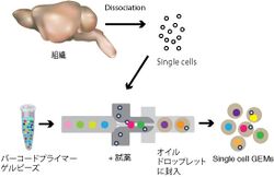
しかしながら、もっとも重要なscRNA-seqの方法論についての進歩は、2015年、Harvard Medical Schoolの独立した2つのグループが、inDrop<ref><pubmed>26000487</pubmed></ref>そしてDrop-seq<ref><pubmed>26000488 </pubmed></ref>という類似した2つの高スループットな方法を開発したことであろう。これらの方法では、[[マイクロ流体力学]] (Microfluidics) 、 UMI(上述)と細胞ごとのCell Barcodeという2種類のDNAバーコーディング、そしてNGSと情報解析を利用している。そして、多く細胞のサンプル調製の自動化と容易さから、1つの細胞あたりに要するコストを大幅に低下させることに成功した(Drop-seqは発表時で、1細胞あたり約5セント)。つまり、細胞1つずつをマイクロ流体力学によるエマルジョン技術を利用した装置に流入させ、その1細胞を1つのDroplet(油中水滴)に自動的に閉じ込める。そのDroplet中には、DropletごとにCell barcode/UMIとしてユニークなDNAバーコードを持つゲルビーズ(Gel Beads in Emulsion, GEMs)が入っており、それを足場に3’末端のみを標的にしたcDNA合成反応を実施することで、同じ細胞に含まれていたmRNAが同じCell barcodeを持つcDNAとして合成され、そのmRNA/cDNAが由来した細胞を識別できるということを利用している(図1)。なお、DropSeqはコストが低いが、細胞の取得率と検出感度が低い弱点がある。inDropはDropSeqより細胞取得率が高く、パラメータを調整することにより、低レベルで発現される遺伝子の検出にも有利であるとされる<ref><pubmed>30472192</pubmed></ref>。 | |||
[http://www.youtube.com/watch?v=fHq9ewdYEWM] | |||
DropSeqのセットアップはDolomite Bio ([https://www.dolomite-bio.com])、inDropは1 Cellbio社から販売されている[https://1cell-bio.com]。しかし、特に重要なのは10x Genomics社が同様の原理を用いた「Chromium」と命名された機器と試薬のシステムを市販することで、多くの研究者に利用できることになったことである[https://www.10xgenomics.com/jp/]。Svenssonらによる最近のデータベース[https://doi.org/10.1101/742304], [http://www.nxn.se/single-cell-studies/gui]では、scRNA-seqを用いた論文で用いられた方法について調査しているが、この数年、10x Genomics社Chromiumを用いた論文が飛躍的に増加し、scRNA-seqの方法として一般的になりつつあることがわかる(現在、10x Genomics社とBioRad社の間で関連特許をめぐる係争がある。)。このシステムは市販であるので導入が容易であり、DropSeqやinDropに比べ多くの転写産物の高感度検出が可能であるが、ランニングコストは高い<ref><pubmed>30472192</pubmed></ref>。なお、3’エンドリード法だけでなく、N末端側に位置する抗体の可変領域などの検出には5’エンドリード法が利用されることがある。 | |||
[[ファイル:ScRNAseqFig1.jpg|サムネイル|250px|'''図1.Droplet使用の3’エンドリード法 '''<br>組織から解離させた細胞それぞれを、マイクロ流体力学を利用した装置で、バーコードプライマーが結合したゲルビーズとともにオイルドロップレットに封じ込める(本文参照)]] | |||
== | ==scRNA-seqの実際== | ||
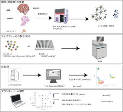
ここでは主流になっている10x Genomics社のChromiumを用いた方法とSMART-seq2などを用いた方法に共通する方法の実際について俯瞰する。scRNA-seqの利用には、4つのステップがある(図2)<ref><pubmed>30089861</pubmed></ref>。1)個体や組織を採集し、そこから細胞あるいは細胞核を個別にすること。2)ChromiumやSMART-seq2などによる個々の細胞からのライブラリーの作製とNGS。3)前処理(preprocessing、得られた配列の整理)。4)ダウンストリーム解析(生物学的な情報を得るコンピューター生物学)。これらのうち、2)の段階については、上に記述したように市販の機器や試薬を利用する機会が多くなっているので、そのためのマニュアル等が参考になるはずである。 | |||
[[ファイル:scRNAseqFig2b.jpg|サムネイル|250px|'''図2.scRNA-seqの実際のステップ '''<br>細胞の単離、ライブラリ作製とNGS、データの前処理から次元圧縮、ダウンストリーム解析。図の一部は2016 DBCLS TogoTV、あるいはR-studio/Seuratを用いて10x Genomics社のPBMCデータ([https://support.10xgenomics.com/single-cell-gene-expression/datasets]から執筆者が作製。]] | |||
[[ファイル: | |||
===組織からの細胞、細胞核の分離=== | ===組織からの細胞、細胞核の分離=== | ||
血液細胞のように浮遊した細胞ではない場合、物理的あるいは酵素処理などによって解離することで、生組織から状態の良いバラバラになった細胞を調製する必要がある。神経系組織の酵素処理には、パパインを用いる方法が広く用いられている。ここで、しばしば問題となるのが、酵素処理のため短時間加温することで、発現量が変化する遺伝子が存在することである<ref><pubmed>27090946</pubmed></ref>。例えば、脳の[[ミクログリア]]の解析には、低温下で組織をホモゲナイズするなどの工夫が必要であった<ref><pubmed>30471926</pubmed></ref>。また、このような現象を抑制するために、酵素処理時に転写阻害剤である[[アクチノマイシン]]で処理したり<ref><pubmed>29024657</pubmed></ref>、ヒマラヤ氷河から得られた細菌Bacillus licheniformisから得られた低温プロテアーゼを用いる方法も報告されている<ref><pubmed>28851704</pubmed></ref>。また、細胞解離後に、メタノールで固定しscRNA-seqに使用することも可能である<ref><pubmed>28526029</pubmed></ref>。 | |||
単離した細胞は、そのまま10x GenomicsのChromiumのプラットフォームに導入することができるが、抗体や蛍光タンパク質レポーターなどを用いたFACS、[[パニング]]、MACS(磁気ビーズカラム)などによる特定のマーカーを細胞表面などに発現する細胞の選択的濃縮や除去を行う場合もある。更に、抗体に抗体表示バーコードDNAをカップリングさせるCITE-seq(Cellular Indexing of Transcriptomes and Epitopes by Sequencing) については、下記のマルチモーダルなオミクスの項目で述べる。 | |||
なお、ヒト組織や希少生物などから生細胞を得ることは困難なことが多い。この場合、scRNA-seqの変法として、凍結した組織から、各細胞由来の核を調製し、核内のmRNAを分析するsnRNA-seq (single-nucleus RNA-seq)が利用されている。ただ、snRNA-seqでは、FACSなどによる特定細胞集団の同定が困難であることが多く、細胞質を持つ生細胞を利用した場合と同等な結果が必ずしも得られない<ref><pubmed>24248345</pubmed></ref><ref><pubmed>26890679</pubmed></ref> <ref><pubmed>27471252</pubmed></ref><ref><pubmed>28846088</pubmed></ref><<ref><pubmed>29220646</pubmed></ref><ref><pubmed>28846088</pubmed></ref><ref><pubmed>30586455</pubmed></ref><ref><pubmed>28729663</pubmed></ref><ref><pubmed>31728515</pubmed></ref><ref><pubmed>32341560</pubmed></ref> [Nature Biotechnology doi: 10.1038/s41587-020-0469-4] 。snRNA-seqでは、組織をそのまま凍結することから開始するので、上述したscRNA-seqの内在的な問題である酵素処理や加温などを避けることができる。こうしたプロトコールの一部は、protocols.ioのHuman Cell Atlasのグループ[https://www.protocols.io/groups/hca]で公開されている。 | |||
更に、RNAを分析するscRNA-seqではないが、遺伝子発現との関係が想定される[[オープンクロマチン]]領域を[[トランスポゾン]]を用いることで個々の細胞レベルで選択的に検出するsingle cell ATAC-seq (Assay for Transposase-Accessible Chromatin)<ref><pubmed>26083756</pubmed></ref>, <ref><pubmed>29434377</pubmed></ref><ref><pubmed>25953818</pubmed></ref>, single cell THS-seq (transposome hypersensitive-site) <ref><pubmed>29227469</pubmed></ref>や[[DNAメチル化]]領域を観察する方法も報告されている<ref><pubmed>28798132</pubmed></ref>。 | |||
===scRNA-seqデータの前処理=== | |||
10x Genomics社のChromium、Illumina社のNGSを利用した場合、Cell Ranger(Linux上で作動)を用いて、各生物種ごとのレファレンス配列リスト([https://www.ncbi.nlm.nih.gov/grc])やEggNOG ([http://eggnogdb.embl.de])などを利用し、細胞とトランスクリプトームの対応マトリックスを作製する。その後のデータの処理についても、10x Genomics社がソフトウェアLoupeを提供している。しかしながら、その後のダウンストリーム解析を考慮して、[[R言語]], [[Python]], MATLABなどのデータ解析のための汎用プログラミング言語やコードで扱えるオブジェクトに変換するのが通常である。ここでは、scRNA-seqデータ解析のために最もよく利用されているR言語を用いたパッケージ「Seurat」<ref><pubmed> 29608179 </pubmed></ref> <ref><pubmed> 31178118 </pubmed></ref>を中心に紹介したい。なお、Pythonを利用したものでは、ドイツ・ミュンヘンInstitute of Computational Biologyの Fabian Theisらが開発しているScanpyが有名である<ref><pubmed> 29409532</pubmed></ref>。 | |||
New York UniversityのRahul Satija研究室が開発しているSeuratは、scRNA-seqデータ解析のために広く利用されているRパッケージであり、2020年4月現在、その最新バージョンはSeurat 3.1.4である。論文の正式発表前から、サポート情報提供やコード修正なども頻繁に行っており、Satija研究室のウェッブサイト( [http://satijalab.org/Seurat])、Github([https://github.com/satijalab/Seurat])、更にTwitterアカウント(@satijalab)などで最新情報を得ることできる。 | |||
最初に行うのは、scRNA-seqデータの品質管理である。ここでは、質の低い細胞のデータ(転写産物の種類が少なく、ミトコンドリア由来の転写産物が多い)を取り除く。また、複数の試料を組み合わせる場合には、バッチごとの違いについて検討する<ref><pubmed>29608177</pubmed></ref>。また、Dropletを使用するscRNA-seqでしばしば問題になるのが、Dropletに2つ以上の細胞が封じ込められ、それらが同一のCell barcodeを持ってしまうアーティファクトである。通常Doubletと呼ばれるこの問題はダウンストリーム解析を混乱させるので、細胞単離の段階から注意する必要があるが、細胞集団に特徴的なマーカー遺伝子が知られていればscRNA-seqデータ取得後にも、データ処理で検討することは可能である。この問題を解決する新たなアプローチも試みられている<ref><pubmed>30954476</pubmed></ref> <ref><pubmed>31836005</pubmed></ref><ref><pubmed>31856883</pubmed></ref> [https://doi.org/10.1101/2019.12.17.879304][https://doi.org/10.1101/699637]。 | |||
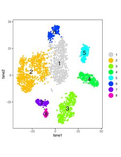
このようなノーマライゼーションの過程を経て、scRNA-seqのデータ解析において、最初に行うのが、[[次元圧縮]] (dimensionality reduction)である<ref><pubmed>30617341</pubmed></ref><ref><pubmed>31780648</pubmed></ref>。PCA (Principal component analysis, 主成分分析)、UMAP(Uniform Manifold Approximation and Projection, 均一マニフォールド近似と投影)、Diffusion maps, t-SNE(t-distributed Stochastic Neighbor Embedding , t分布型確率的近傍埋込み)などの手法が用いられる。 特に、t-SNE[http://www.jmlr.org/papers/v9/vandermaaten08a.html]は、高次元データを低次元の点の集合として可視化することで、それぞれの細胞の持つトランスクリプトームの類似度についての直観的な表示が可能でありしばしば用いられる(図3)。次に、[[Louvainアルゴリズム]]などでクラスタリング(コミュニティ分割)を行いグラフ上に表示できる(図3の色分け)。こうして、違ったタイプの細胞の集合が別のクラスターとして表示される。しかし、データによっては、tSNEだけでなく、他の方法でパラメータの調節を行うことで違ったデータ解釈(別の細胞クラスターの同定)ができるケースもあり、違った方法も試してみることが推奨される。 | |||
[[ファイル:ScRNAseqFig3.jpg|サムネイル|250px|'''図3.t-SNE によるデータ可視化'''<br>ニワトリ網膜の視細胞のデータを用いて作製[A]。]] | |||
==ダウンストリーム解析== | |||
===細胞クラスターの解釈とマーカー遺伝子候補の発見=== | |||
scRNA-seqデータから得られる生物学的知見には、内在的に存在する細胞の種類、外部刺激や環境で変化した細胞の状態、そして種類や変化により特徴的に発現するマーカー遺伝子候補の発見がある<ref><pubmed>27824854</pubmed></ref><ref><pubmed>32033589</pubmed></ref> | |||
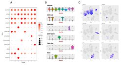
。クラスタリングにより、異なった細胞集団の存在が認識されると、それぞれのクラスターに特徴的に発現している遺伝子を具体的に探索し、細胞集団の持つバイオマーカーによって、そのクラスターの同定が可能になる。例えば、既に神経細胞とグリア細胞に特異的に発現する典型的マーカーはよく知られており、それぞれのクラスターの識別は容易である。更に、神経細胞のタイプを区別できるマーカーや、外部刺激によって遺伝子発現が変化した神経細胞の状態は、In situ hybridizationや免疫組織化学などにより確認できる。このようなクラスターごとに発現が異なる遺伝子(差次的発現遺伝子)を見つけるためには(Differential expression analysis, DE analysis)、SeuratのFindMarkersコマンド中でも利用可能である専用コード(MAST <ref><pubmed>26653891</pubmed></ref>、DESeq2 <ref><pubmed>25516281</pubmed></ref>など)を用いることができる。scRNA-seqの解析に必要なコードは、scRNA-tools [https://www.scrna-tools.org], Awesome single cell [https://github.com/seandavi/awesome-single-cell], Bioconductor[https://www.bioconductor.org]で紹介されており、ほとんどがダウンロード可能である。また、最新の情報については、bioRxivなどのプレプリントサーバで公開されていることが多く、scRNA-seqのデータ(下記参考)とともに、オープンサイエンス実践の好例となっている。細胞ごとの差次的発現遺伝子の可視化には、ドットプロット(dot plot)、ヴァイオリンプロット(violin plot)、次元圧縮の可視化図に重ねる布置プロット(feature plot)などが頻繁に用いられる(図4)。 | |||
[[ファイル:ScRNAseqFig4.jpg|サムネイル|250px|'''図3.scRNA-seqデータの可視化の例 '''<br>A. ドットプロット。B.ヴァイオリンプロット。C. 次元圧縮と重ねた布置プロット。ニワトリ網膜の視細胞のデータを用いて作製[A]。]] | |||
=== | ===偽時系列解析、遺伝子制御ネットワーク、パスウェイ解析=== | ||
実験的なノイズとは別に生物学的に意味のある遺伝子発現の変動には、位置情報、[[細胞周期]]、[[概日リズム]]、発現変動が大きい破裂型プロモーターの作動などの理由で変動が見られるものもある<ref><pubmed> 31217225 </pubmed></ref><ref><pubmed> 26000846</pubmed></ref>。特に、刺激・薬剤処理やさまざまな病態の進行や治療に伴う細胞の変化、発生途上の細胞系譜や細胞分化といった細胞の遷移状態の解析([[偽時系列解析]]Pseudo-time analysis )には、scRNA-seqデータを用いることが効果的である<ref><pubmed>29576429</pubmed></ref><ref><pubmed>28813177</pubmed></ref><ref><pubmed>29565398</pubmed></ref>。これらの分析のためには[[軌道推定]](Trajectory inference)の解析手法が用いられる。しばしば用いられるMonocle3 <ref><pubmed>30787437</pubmed></ref>など、多くのコードを収集しているGithubのサイトがある [https://github.com/dynverse/dynmethods][https://github.com/agitter/single-cell-pseudotime]。RNA velocityといった転写産物のスプライシングの状態から細胞の分化状態を推定する方法もある<ref><pubmed>30089906</pubmed></ref>。しかし、これらの方法は、あくまで発生途上の[[細胞系譜]]や細胞分化の推定に過ぎない。細胞系譜を更に確実に観察しつつ、scRNA-seqを行うことで、細胞タイプの系統関係を調べる方法として、CRISPR-Cas9を用いた[[ゲノム編集]]による記録法を導入したscGESTALT<ref><pubmed>29608178</pubmed></ref>、ScarTrace<ref><pubmed>29590089</pubmed></ref> 、LINNAEUS<ref><pubmed>29644996</pubmed></ref>がある。 | |||
• また細胞分化や変動に伴う特徴的な遺伝子発現をscRNA-seqで観察することは、遺伝子制御ネットワーク(例、SCENIC<ref><pubmed>28991892</pubmed></ref>, [https://github.com/aertslab/SCENIC])や[[代謝経路]]や[[シグナル伝達系]]のための[[パスウェイ解析]](例、Metascape<ref><pubmed>30944313</pubmed></ref>, [http://metascape.org])を理解するシステム生物学的な研究として有用である。更に、scRNA-seqで得られた結果をもとに、細胞間相互作用の理解を深めるのを目的とするCellPhoneDB<ref><pubmed>32103204</pubmed></ref>[https://github.com/Teichlab/cellphonedb]、NicheNet<ref><pubmed>3181926</pubmed></ref>, SVCA<ref><pubmed>31577949</pubmed></ref>がある。Perturb-seq<ref><pubmed>27984732</pubmed></ref> やその変法<ref><pubmed> 32231336</pubmed></ref>は、CRISPRライブラリーによるゲノム編集を施した細胞をscRNA-seqで解析することで、遺伝子機能や遺伝子間の相互作用の理解を可能にしている。 | |||
== | ==scRNA-seqの神経科学研究への適用== | ||
===神経系細胞ビッグデータとしてのscRNA-seq=== | |||
様々な神経・精神疾患について理解しその診断や治療に役立てるためには、神経細胞、[[グリア細胞]]を中心にした神経系にある細胞の種類や状態を識別し、それぞれの細胞における分子的な変化を観察することが重要である <ref><pubmed>28775344</pubmed></ref><ref><pubmed>29738987</pubmed></ref>。本本項目で解説してきたscRNA-seqの技術は、神経系に見られるそれぞれの細胞のトランスクリプトームについて[[ビッグデータ]]を提供することで、この細胞の種類や状態の識別に新たな根拠を与えつつある。近年、中枢神経系の[[アストロサイト]]、[[オリゴデンドロサイト]]、ミクログリアといったグリア細胞も均一ではなく、内在的な多様性や外部因子による状態の変動が報告されてきている。神経細胞は、著しく多様であり、この多様性が神経系を特徴づけており、その多彩で複雑な機能の発現に必須である。従来の神経科学では、神経細胞の多様性は、それぞれの神経細胞の解剖学的な位置、発現している分子、電気生理学、結合性、形態、神経伝達物質、神経伝達物質受容体とシグナル伝達によって識別されてきている。こうした神経細胞の多様性を便宜的に記述するのに、タイプ(type)、クラス(class)、サブクラス(subclass)、サブタイプ(subtype) というような用語が用いられてきた。しかし、ここでは混乱を防ぐため、Masland(2004)<ref><pubmed>15242626</pubmed></ref>が提唱し、広く受けいれられている「クラス」と「タイプ」という単語を用いることとする。タイプは、これ以上分類することができないとされる階層であり、共通性を持つタイプの集団がクラスである。例えば、大脳皮質の錐体細胞、網膜神経節細胞といった大雑把な区分はクラスである。大脳皮質の錐体細胞というクラスは、層や領野によって異なるタイプ、網膜神経節細胞には視覚情報に対して応答が異なるタイプが存在する。scRNA-seqは、「タイプ」の理解に新たな視点を提供している。 | |||
=== | |||
===神経系へのscRNA-seqの適用=== | ===神経系へのscRNA-seqの適用=== | ||
[[大脳皮質]]には、[[錐体細胞]]や[[非錐体細胞]]などの神経細胞や様々なグリア細胞などが見られ、古くから神経細胞タイプの識別が行われてきた。初期のscRNA-seq技術でも、マウス皮質の小規模な細胞数を分類した研究で、これまで知られていた主要な細胞タイプとは違うタイプが見つかりその有効性が示された<ref><pubmed>25700174</pubmed></ref>。その後のDroplet使用の3’エンドリード法を利用した多数の細胞数の解析で、更に多数の神経細胞のタイプが見つかっている<ref><pubmed>28846088</pubmed></ref><ref><pubmed>30096299</pubmed></ref><ref><pubmed>30096314</pubmed></ref><ref><pubmed>30382198</pubmed></ref><ref><pubmed>29320739</pubmed></ref><ref><pubmed>28846088</pubmed></ref>[https://doi.org/10.1101/2020.03.14.991018]。特に、GABA作動性介在神経細胞タイプの多様性とその発生<ref><pubmed>28942923</pubmed></ref><ref><pubmed>28134272</pubmed></ref><ref><pubmed>29472441</pubmed></ref><ref><pubmed>29513653</pubmed></ref>についての情報は重要であろう。また、初期の発生過程<ref><pubmed>26940868</pubmed></ref><ref><pubmed>30485812</pubmed></ref><pubmed>31073041</pubmed></ref><ref><pubmed>30635555</pubmed></ref><ref><pubmed>30625322</pubmed></ref>、老化<ref><pubmed>31551601</pubmed></ref><の理解が、scRNA-Seq技術を利用することで進んでいる。更に、[[神経活動]]によって変化するトランスクリプトームの変化も細胞ごとに調査され興味深い<ref><pubmed>29230054</pubmed></ref> 。 ヒトを含めた霊長類の大脳についても発達段階を含めてscRNA-seqが適用されてきている<ref><pubmed>26060301</pubmed></ref><ref><pubmed>27339989</pubmed></ref><ref><pubmed>29539641</pubmed></ref><ref><pubmed>29217575</pubmed></ref><ref><pubmed>28846088</pubmed></ref><ref><pubmed>29227469</pubmed></ref><ref><pubmed>31303374</pubmed></ref><ref><pubmed>29867213</pubmed></ref><ref><pubmed>31435019</pubmed></ref><ref><pubmed>32424074</pubmed></ref> [https://doi.org/10.1101/709501[https://doi.org/10.1101/2020.03.31.016972][https://doi.org/10.1101/2020.04.23.056390]。ヒトや霊長類に特徴的とされる[[島]]のvon Economo神経細胞(紡錘細胞)のような希少な神経細胞のscRNA-seqにも成功している<ref><pubmed>32127543</pubmed></ref>。 | |||
[[海馬]]<ref><pubmed>29241552</pubmed></ref><ref><pubmed>29912866</pubmed></ref><ref><pubmed>29335606</pubmed></ref><ref><pubmed>31942070</pubmed></ref>では、これまでの研究で記載されてきた神経細胞のタイプの存在が確認され、更に新規のタイプが見つかった。中枢神経系では、その他、[[外側膝状体]]<ref><pubmed>29343640</pubmed></ref>、[[大脳基底核]](足底核)<ref><pubmed>28384468</pubmed></ref> 、[[視床下部]]<ref><pubmed>28166221</pubmed></ref><ref><pubmed>28355573</pubmed></ref> <ref><pubmed>27991900</pubmed></ref><ref><pubmed>30385464</pubmed></ref> <ref><pubmed>31249056</pubmed></ref><ref><pubmed>30858605</pubmed></ref>、[[線条体]]<ref><pubmed>27425622</pubmed></ref><ref><pubmed>30134177</pubmed></ref><ref><pubmed>31875543</pubmed></ref>、[[中脳]]<ref><pubmed>27716510</pubmed></ref><ref><pubmed>29499164</pubmed></ref> <ref><pubmed>30718509</pubmed></ref> 、[[手綱]]<ref><pubmed>29576475</pubmed></ref>、発生中の[[間脳]]<ref><pubmed>30872278</pubmed></ref> 、さらに[[小脳]]<ref><pubmed>30220501</pubmed></ref><ref><pubmed>30735127</pubmed></ref><ref><pubmed>30690467</pubmed></ref>などの結果が得られている。マウスの小脳においては、分子層にこれまでの星状細胞、バスケット細胞というカテゴリーとは違った2種類の神経細胞があることが示唆されている[https://doi.org/10.1101/2020.03.04.976407]。 | |||
脳の外部では、[[運動神経]][ https://doi.org/10.1101/2020.03.16.992958]、[[感覚神経]]<ref><pubmed>25420068</pubmed></ref><ref><pubmed>26691752</pubmed></ref>、[[らせん神経節]]<ref><pubmed>30078709</pubmed></ref><ref><pubmed>30209249</pubmed></ref> 、[[臭覚神経]]<ref><pubmed>26541607</pubmed></ref> <ref><pubmed>32059767</pubmed></ref>、[[腸神経系]] <ref><pubmed>29483303</pubmed></ref>[https://doi.org/10.1101/2020.03.02.955757] 、[[網膜]]<ref><pubmed>27565351</pubmed></ref><ref><pubmed>29909983</pubmed></ref><ref><pubmed>30018341</pubmed></ref><ref><pubmed>31260032</pubmed></ref><ref><pubmed>31128945</pubmed></ref><ref><pubmed>30712875</pubmed></ref><ref><pubmed>30548510</pubmed></ref><ref><pubmed>31075224</pubmed></ref><ref><pubmed>31399471</pubmed></ref><ref><pubmed>31848347</pubmed></ref><ref><pubmed>31673015</pubmed></ref><ref><pubmed>31653841</pubmed></ref><ref><pubmed>31784286</pubmed></ref>[https://doi.org/10.1101/2020.02.26.966093][ https://doi.org/10.1101/779694][https://www.biorxiv.org/content/10.1101/617555][A]でのscRNA-seqデータがある。また[[iPS細胞]]や[[ES細胞]]由来の神経組織[[オルガノイド]]に含まれる神経細胞タイプを知る上でも利用されている<ref><pubmed>28094016</pubmed></ref><ref><pubmed>28279351</pubmed></ref><ref><pubmed>31168097</pubmed></ref><ref><pubmed>31996853</pubmed></ref><ref><pubmed>31968264</pubmed></ref><ref><pubmed><ref><pubmed>32221280</pubmed></ref></pubmed></ref>。 | |||
===神経細胞以外の細胞=== | |||
[[上衣細胞]]<ref><pubmed>29727663</pubmed></ref>は、[[神経幹細胞]]としての役割が示唆されてきたが、scRNA-seqによる解析ではその可能性が支持されなかった。グリア細胞では、[[ラジアルグリア]]<ref><pubmed>26406371</pubmed></ref><ref><pubmed>25734491</pubmed></ref><ref><pubmed>29281841</pubmed></ref><ref><pubmed>29217575</pubmed></ref><ref><pubmed>29539641</pubmed></ref>[A]、アストロサイト<ref><pubmed>32139688</pubmed></ref><ref><pubmed>32203496</pubmed></ref>[ https://doi.org/10.1101/2020.04.27.064881]に多様性があることが示唆されてきている。また、オリゴデンドロサイト<ref><pubmed>27284195</pubmed></ref><ref><pubmed>30078729</pubmed></ref><ref><pubmed>30257220</pubmed></ref><ref><pubmed>30747918</pubmed></ref><ref><pubmed>31958186</pubmed></ref>[https://doi.org/10.1101/2020.03.06.981373] [A]については、これまで細胞生物学的に研究されてきた分化の過程がscRNA-seqにより検出されている。ミクログリアは、神経系の発達、老化、損傷などに伴うトランスクリプトームの変化がscRNA-seqにより詳細に明らかになった<ref><pubmed>27338705</pubmed></ref><ref><pubmed>29020624</pubmed></ref><ref><pubmed>30206190</pubmed></ref><ref><pubmed>30471926</pubmed></ref><ref><pubmed>31209379</pubmed></ref> | |||
<ref><pubmed>31835035</pubmed></ref>。また、[[CNS境界関連マクロファージ]](BAM) <ref><pubmed>31061494</pubmed></ref>や[[脳血管系]]<ref><pubmed>29443965</pubmed></ref>のscRNA-seqも実施されている。 | |||
===疾患=== | |||
scRNA-seqは、疾患の理解にも有用である。筋萎縮性側索硬化症<ref><pubmed>30948552</pubmed></ref>、多発性硬化症<ref><pubmed>30747918</pubmed></ref><ref><pubmed>30420755</pubmed></ref><ref><pubmed>32313246</pubmed></ref> | |||
、アルツハイマー病やそのモデル動物<ref><pubmed>31042697</pubmed></ref><ref><pubmed>31399126</pubmed></ref><ref><pubmed>29020624</pubmed></ref> | |||
[https://doi.org/10.1101/628347]<ref><pubmed>28602351</pubmed></ref><ref><pubmed>32341542</pubmed></ref>、統合失調症<ref><pubmed>29785013</pubmed></ref><ref><pubmed>32203495</pubmed></ref>、自閉症やレット症候<ref><pubmed>31097668</pubmed></ref><ref><pubmed>30455458</pubmed></ref> | |||
、シャルコー・マリー・トゥース病<ref><pubmed>29888333</pubmed></ref>、ダウン症[https://www.biorxiv.org/content/10.1101/2020.01.01.892398v1]、パーキンソン病<ref><pubmed>30503143</pubmed></ref>[https://doi.org/10.1101/2020.04.29.067587]、がん<ref><pubmed>31327527</pubmed></ref><ref><pubmed>28360267</pubmed></ref>などに適用されている。 | |||
==scRNA-seqの展望== | ==scRNA-seqの展望== | ||
===神経系の多様性と進化=== | ===神経系の多様性と進化=== | ||
NGSを用いることで、どんな生物種にも適用可能なscRNA-seqは、既に多様な生物の神経系の細胞の理解、更には種間の相同性や差異の研究に利用されており、神経系の進化を細胞レベルで考察するのに有用であろう(例、センチュウ<ref><pubmed> 28818938 </pubmed></ref>、ショウジョウバエ <ref><pubmed>29909982</pubmed></ref><ref><pubmed>29149607</pubmed></ref><ref><pubmed>30703584</pubmed></ref><ref><pubmed>29909983</pubmed></ref>、カタユウレイボヤCiona intestinalis <ref><pubmed>30069052</pubmed></ref><ref><pubmed>30228204</pubmed></ref>、ゼブラフィッシュ<ref><pubmed>31018142</pubmed></ref><ref><pubmed>30929901</pubmed></ref>、アカミミガメTrachemys scripta elegans、トカゲPogona vitticeps, pv<ref><pubmed>29724907</pubmed></ref>、ニワトリ[A]、霊長類<ref><pubmed>30730291</pubmed></ref><ref><pubmed>31619793</pubmed></ref>[https://doi.org/10.1101/2020.03.31.016972])。ただ、遺伝子やトランスクリプトームの研究が進んでいる生物種では比較的容易であるが、遺伝子のアノテーションが十分でない生物種を用いる場合、scRNA-seqのデータ解析は困難を伴うので、NCBIのTaxonomy[https://www.ncbi.nlm.nih.gov/taxonomy]やEggNOG [http://eggnogdb.embl.de] <ref><pubmed>30418610</pubmed></ref>などを利用する。また種を超えた細胞タイプの相同性の理解には様々な工夫が必要である<ref><pubmed>31552245</pubmed></ref><ref><pubmed>30712875</pubmed></ref> | |||
===データベースと統合=== | ===データベースと統合=== | ||
獲得されたscRNA-seqのデータは様々な目的で利用できるので、データベース化し利用できるようにする必要がある。神経系のトランスクリプトーム一般のデータベースが多数公開されており<ref><pubmed>29437890</pubmed></ref>、scRNA-seqのデータも基本的にNCBIのGene Expression Omnibus[https://www.ncbi.nlm.nih.gov/geo/]に登録されている。また、オープンサイエンス推進のためにcommon coordinate framework (CCF) やcentral annotation platform (CAP)という概念のもと、特にscRNA-seqを意識したものとして、米国のBRAIN Initiative Cell Census Consortium<ref><pubmed>29096072</pubmed></ref> 、Human Cell Atlas ProjectのHuman Cell Atlas Data Portal [https://data.humancellatlas.org]、アレン脳研究所のAllen Brain Atlas [https://portal.brain-map.org]、ブロード研究所のSingle Cell Portal [https://singlecell.broadinstitute.org/]などのデータベースが稼働している。また、異なった方法や実験で得られたscRNA-seqのデータを体系的に比較することも重要であり、Seurat 3.0以降に組み込まれたMMN (Mutual Nearest Neighbors)、LIGER<ref><pubmed>31178122</pubmed></ref> 、Harmony<ref><pubmed>31740819</pubmed></ref> 、MetaNeighber<ref><pubmed>29491377</pubmed></ref>のようなアルゴリズムが開発されている。 | |||
===空間トランスクリプトミクス=== | ===空間トランスクリプトミクス=== | ||
多数の細胞を扱うscRNA-seqの弱点は、組織から細胞や細胞核を解離する必要があるので、その細胞が存在していた解剖学的あるいは空間的な位置の情報を消去してしまうということである。組織切片におけるタンパク質などの分布は免疫組織化学、mRNAの分布はin situ hybridizationで検出することができるが、数多くのmRNAの分布を情報処理技術と組み合わせ一気に同定する方法がscRNA-seqと同様に開発されてきている。Slide-seq<ref><pubmed>30923225</pubmed></ref>[ https://doi.org/10.1101/2020.03.12.989806]、osmFISH<ref><pubmed>30377364</pubmed></ref>、STARmap (spatially-resolved transcript amplicon readout mapping), <ref><pubmed>29930089</pubmed></ref>、seqFISH <ref><pubmed>27764670</pubmed></ref>、pciSeq(probabilistic cell typing by in situ sequencing)、DSP(Digital Spatial Profiling) <ref><pubmed>32393914</pubmed></ref>、Expansion sequencing[http://doi.org/10.1101/2020.05.13.094268] | |||
、更に10x Genomics社が市販するVisiumなどがある。現状では、大きな組織の空間トランスクリプトミクスは、空間解像度が細胞レベルにいたっておらず、技術普及の観点からも課題が多い。しかし、そのデータを解析するためのアルゴリズム<ref><pubmed>29553578</pubmed></ref><ref><pubmed>29553579</pubmed></ref><ref><pubmed>32350282</pubmed></ref> [https://doi.org/10.1101/757096][ https://doi.org/10.1101/701680] [https://doi.org/10.1101/431957]や、更にMerFish <ref><pubmed>25858977</pubmed></ref>、corrFISH <ref><pubmed>27271198</pubmed></ref>のように、subcellularレベルで多数のmRNAを検出する方法が開発されてきており、scRNA-seqと組み合わせることで、その弱点を補う空間トランスクリプトミクスにも利用され始め<ref><pubmed>30385464</pubmed></ref>、今後の発展が期待される。 | |||
===マルチモーダルなシングルセルオミクス=== | |||
同一の細胞からscRNA-seqの情報だけでなく、ゲノム配列、ATAC-seqなどによるエピゲノム解析、少数のタンパク質、あるいはプロテオームなどを、同時に記録するマルチモーダルなオミクスが注目されている<ref><pubmed>31907462</pubmed></ref><ref><pubmed>30696980</pubmed></ref>。2019年には、Nature Methodsの「Methods of the Year」に選ばれており、現状については、その特集号などを参考にされたい。 | |||
マルチモーダルなシングルセルオミクスとして、神経科学分野で注目されるのは、scRNA-seqをパッチクランプによる電気生理学的情報と組み合わせたPatch-seq<ref><pubmed>26689544</pubmed></ref> <ref><pubmed>26689543</pubmed></ref>である。また、細胞表面分子に対する抗体にDNAを付加することで、マーカーを発現する細胞のトランスクリプトームを観察するCITE-seq<ref><pubmed>28759029</pubmed></ref>、 REAP-seq<ref><pubmed>28854175</pubmed></ref>は既知の細胞マーカーの発現とscRNA-seqが同時に観察できるマルチモーダルなオミクスである。また、BARseq (barcoded anatomy resolved by sequencing) <ref><pubmed>31626774</pubmed></ref>[https://doi.org/10.1101/378760 | |||
]のような方法は、コネクトーム(神経細胞の結合性)と遺伝子発現を記録できるオミクスの新たな方向として興味深い。https://doi.org/10.1101/378760 | |||
== 関連項目 == | == 関連項目 == | ||
*[[ゲノムワイド関連解析 ]] | *[[ゲノムワイド関連解析 ]] | ||
| 132行目: | 115行目: | ||
*[[免疫組織化学法]] | *[[免疫組織化学法]] | ||
*[[エピジェネティクス]] | *[[エピジェネティクス]] | ||
== 参考文献 == | == 参考文献 == | ||
<references /> | <references /> | ||
2020年5月27日 (水) 10:58時点における版
山形方人
Harvard University
DOI:10.14931/bsd.8038 原稿受付日:年月日 原稿完成日:年月日
担当編集委員:
英:single cell RNA sequencing, scRNA-seq
シングルセルRNAシーケンシング(single cell RNA sequencing, 以下scRNA-seq)は、次世代シーケンシング (next generation sequencing、以下NGS)技術を使用して個々の細胞が発現しているmRNA全体、つまりトランスクリプトームを質的、量的に網羅的に調べ、細胞ごとの違いを高解像度で検出、分類することで、細胞の分類を行うことができる分子生物学的、コンピュータ生物学的技術である。また、刺激、発生など細胞の状況に応じて、個々の細胞のトランスクリプトームの情報を得ることで、病態や細胞系譜などの解析も可能である。特に多様な神経細胞が存在する神経系では、この方法により、神経細胞や非神経細胞の分類や状態についての知見が深まり、更に新しいバイオマーカー(biomarker)の発見などが系統的かつ網羅的に行われるようになった。
scRNA-seqとその開発史
トランスクリプトーム
トランスクリプトーム(transcriptome)は、細胞中に存在する全ての転写産物(タンパク質をコードするmRNA、タンパク質をコードしないノンコーディングRNA、マイクロRNAなど)の総体である[1][2]。トランスクリプトームは、ゲノムとは異なり、同一の個体でも、組織ごとに、更には発生段階や細胞外環境や刺激によって変化する。トランスクリプトームは、同質あるいは異質の多数の細胞集団(組織、培養細胞)からRNA抽出後、cDNAに変換し、それを1990年代に出現したDNAマイクロアレイのように数多くの既知mRNAを識別する技術によって解析されるようになった。その後、NGSの利用により、希少mRNAやノンコーディングRNAを含めた未知の転写産物の高感度検出が可能になるとともに、スプライシングで成熟していく過程のmRNAなど、転写産物の種類だけでなく、転写産物の構造的差異(スプライシングバリアント、SNPs、変異など)の解析もできるようになった。加えて、ヒトやモデル実験生物(マウス、ゼブラフィッシュ、ショウジョウバエ、センチュウなど)だけでなく、多種多様な生物のトランスクリプトームの把握も可能になった。ここでは、このような多数の細胞集団、つまり個体や特定の組織全体ではなく、細胞1つの持つトランスクリプトームを解析する方法(scRNA-seq)とそのscRNA-seqデータを利用することで得られる情報について概説する。
scRNA-seqとその開発史
1つの細胞の持つ生体物質を解明し、定量しようとする試みは古くからあった。1960年代になると、フローサイトメトリーを利用した蛍光活性化セルソーティング(Fluorescence-activated cell sorting, FACS)が発明され、標識抗体などのプローブと組み合わせることで、多数の細胞集団の中で1つの細胞が持っている分子の種類や量についての断片的な研究が可能になり、この方法は現在でも汎用されている[3]。その後、免疫組織化学やin situ hybridizationnなどにより、タンパク質やmRNAの種類や量が観察できるようになり、組織中に存在するそれぞれの細胞の同定などに活用されてきている。最近では、それぞれの細胞が持つ抗原分子を、異なった金属イオンで標識した抗体とフローサイトメトリーを組み合わせた方法で検出するマスサイトメトリー(CyTOFなど)も開発されてきている[4]。
1つの細胞内にある全RNA(ribosomal RNAを含む)は細胞種にもよるが1-50pgである。そのうち、mRNAの占める割合は1-5%程度である[5]。この微量のmRNAをcDNAに変換してから大幅に増幅できる方法が発明されたことで、1つの細胞が発現するmRNAを高感度で検出できるようになった[6][7] 。例えば、1991年、Linda BuckとRichard Axelは、嗅覚受容体がGタンパク質であると仮定し、個々の嗅覚細胞で特異的に観察されるGタンパク質mRNAを比較することで、嗅覚受容体の同定に成功した[8]。1995年になると、Catherine DulacとRichard Axelは、異なる鋤鼻神経細胞で特異的に発現する遺伝子を1つの細胞から作製したcDNAライブラリーを比較するディファレンシャル・スクリーニングを行うことで、フェロモン受容体を同定した[9]。同じ手法で異なる種類の神経細胞で発現している遺伝子も同定され[10][11]、1つの細胞の持つトランスクリプトームを比較するアプローチが神経細胞で特徴的に発現している遺伝子の同定に効果的なことを示した。
一方で多くの種類のmRNAを1細胞レベルで一挙に観察するトランスクリプトームのための技術にはブレークスルーが待たれた。1つの問題は多種類のcDNAを簡便に識別することを可能にする方法の開発であった。これを可能にしたのが、PCRなどのcDNA増幅法の改良とマイクロアレイの利用であった[12][13]。しかしながら、細胞ごとに高価なマイクロアレイを使用することは、多数の細胞のトランスクリプトームの観察には限界があった。2009年になると、これらの問題を解決できる可能性として、NGSを利用するscRNA-seqプロトコールがAzim Suraniのグループによって報告された[14]。しかしながら、この報告でもわずか8個の細胞の解析に留まっており、1つの細胞ごとに処理を行うという操作が必要で、多数の細胞についてのトランスクリプームを一挙に理解することはできなかった。また、塩基配列の違うcDNAごとにPCR効率に差がある結果生じる増幅バイアス、また3’末端側が選択的に補足されることなどの課題を残した。
scRNA-seqの現状
その後、5’末端側も含めた完全長cDNAを増幅するscRNA-seqのプロトコールが考案された。特に、SMART-seq(Switch mechanism at the 5' End of RNA Templates)[15]およびその改良されたプロトコールであるSMART-seq2[16] [17]の使用例が多い(既に、SMART-seq3という改良プロトコールもある[2] Single-cell RNA counting at allele and isoform resolution using Smart-seq3 )。また、類似法としてSTRT(single-cell tagged reverse transcription)[18]がある。更に、MARS-seq(Massively parallel single-cell RNA-seq)[19]、CEL-seq(Cell Expression by Linear amplification and Sequencing)[20]、CEL-seq2[21]では、T7 RNAポリメラーゼによるin vitro転写を用いることにより、PCRで顕著な増幅バイアスを減少させようとしている。
増幅バイアス除去のアプローチとして特に重要なのは、2011年に発表された分子識別子(unique molecular identifiers: UMI)を持つcDNAを増幅させ、NGS後の情報処理を用いる方法である[22]。この方法では逆転写反応の際、ランダムなUMIをcDNA末端に付加した後、増幅反応、NGSを行い、cDNA配列とUMI配列の両方を読む。同一のUMIを持っていれば、逆転写時に同一のcDNA由来とカウントする。UMIをカウントすることで、増幅前のmRNAのコピー数を知ることができる[23][24][25] [26][27][28]。2013年には、このような1細胞のシーケンシング技術が、Nature Methods誌のMethod of the Year に選ばれた[3]。
これらの方法のうち、SMART-seq、その改良法であるSMART-seq2は、マイクロピペットによる捕獲、セルソーター、レーザー捕獲などを用いるマルチウェル法、あるいは半導体集積回路製作技術で作った流体集積回路を利用するFluidigm C1の装置[4]と組み合わせることで利用される機会が多い[29][5]。このSMART-seq2プロトコールの特徴は、全長トランスクリプトームを得ることができることであり、mRNAのスプライシングバリアントなどのアイソフォーム、アリルごとの発現情報が得られるSNPs、変異の検出にも利用できる。また、それぞれ細胞ごとの反応を独立した場所で行うため、別の細胞の反応と混じる可能性がない。これらの点が、次に説明するDropletを使用して3’末端のみを標的にしたscRNA-seqに比べた場合の長所であるが、その高コスト(1細胞あたり数十ドル)と処理可能な細胞数の少なさが短所である。
Droplet使用の3’エンドリード法
しかしながら、もっとも重要なscRNA-seqの方法論についての進歩は、2015年、Harvard Medical Schoolの独立した2つのグループが、inDrop[30]そしてDrop-seq[31]という類似した2つの高スループットな方法を開発したことであろう。これらの方法では、マイクロ流体力学 (Microfluidics) 、 UMI(上述)と細胞ごとのCell Barcodeという2種類のDNAバーコーディング、そしてNGSと情報解析を利用している。そして、多く細胞のサンプル調製の自動化と容易さから、1つの細胞あたりに要するコストを大幅に低下させることに成功した(Drop-seqは発表時で、1細胞あたり約5セント)。つまり、細胞1つずつをマイクロ流体力学によるエマルジョン技術を利用した装置に流入させ、その1細胞を1つのDroplet(油中水滴)に自動的に閉じ込める。そのDroplet中には、DropletごとにCell barcode/UMIとしてユニークなDNAバーコードを持つゲルビーズ(Gel Beads in Emulsion, GEMs)が入っており、それを足場に3’末端のみを標的にしたcDNA合成反応を実施することで、同じ細胞に含まれていたmRNAが同じCell barcodeを持つcDNAとして合成され、そのmRNA/cDNAが由来した細胞を識別できるということを利用している(図1)。なお、DropSeqはコストが低いが、細胞の取得率と検出感度が低い弱点がある。inDropはDropSeqより細胞取得率が高く、パラメータを調整することにより、低レベルで発現される遺伝子の検出にも有利であるとされる[32]。 [6] DropSeqのセットアップはDolomite Bio ([7])、inDropは1 Cellbio社から販売されている[8]。しかし、特に重要なのは10x Genomics社が同様の原理を用いた「Chromium」と命名された機器と試薬のシステムを市販することで、多くの研究者に利用できることになったことである[9]。Svenssonらによる最近のデータベース[10], [11]では、scRNA-seqを用いた論文で用いられた方法について調査しているが、この数年、10x Genomics社Chromiumを用いた論文が飛躍的に増加し、scRNA-seqの方法として一般的になりつつあることがわかる(現在、10x Genomics社とBioRad社の間で関連特許をめぐる係争がある。)。このシステムは市販であるので導入が容易であり、DropSeqやinDropに比べ多くの転写産物の高感度検出が可能であるが、ランニングコストは高い[33]。なお、3’エンドリード法だけでなく、N末端側に位置する抗体の可変領域などの検出には5’エンドリード法が利用されることがある。

組織から解離させた細胞それぞれを、マイクロ流体力学を利用した装置で、バーコードプライマーが結合したゲルビーズとともにオイルドロップレットに封じ込める(本文参照)
scRNA-seqの実際
ここでは主流になっている10x Genomics社のChromiumを用いた方法とSMART-seq2などを用いた方法に共通する方法の実際について俯瞰する。scRNA-seqの利用には、4つのステップがある(図2)[34]。1)個体や組織を採集し、そこから細胞あるいは細胞核を個別にすること。2)ChromiumやSMART-seq2などによる個々の細胞からのライブラリーの作製とNGS。3)前処理(preprocessing、得られた配列の整理)。4)ダウンストリーム解析(生物学的な情報を得るコンピューター生物学)。これらのうち、2)の段階については、上に記述したように市販の機器や試薬を利用する機会が多くなっているので、そのためのマニュアル等が参考になるはずである。

細胞の単離、ライブラリ作製とNGS、データの前処理から次元圧縮、ダウンストリーム解析。図の一部は2016 DBCLS TogoTV、あるいはR-studio/Seuratを用いて10x Genomics社のPBMCデータ([1]から執筆者が作製。
組織からの細胞、細胞核の分離
血液細胞のように浮遊した細胞ではない場合、物理的あるいは酵素処理などによって解離することで、生組織から状態の良いバラバラになった細胞を調製する必要がある。神経系組織の酵素処理には、パパインを用いる方法が広く用いられている。ここで、しばしば問題となるのが、酵素処理のため短時間加温することで、発現量が変化する遺伝子が存在することである[35]。例えば、脳のミクログリアの解析には、低温下で組織をホモゲナイズするなどの工夫が必要であった[36]。また、このような現象を抑制するために、酵素処理時に転写阻害剤であるアクチノマイシンで処理したり[37]、ヒマラヤ氷河から得られた細菌Bacillus licheniformisから得られた低温プロテアーゼを用いる方法も報告されている[38]。また、細胞解離後に、メタノールで固定しscRNA-seqに使用することも可能である[39]。
単離した細胞は、そのまま10x GenomicsのChromiumのプラットフォームに導入することができるが、抗体や蛍光タンパク質レポーターなどを用いたFACS、パニング、MACS(磁気ビーズカラム)などによる特定のマーカーを細胞表面などに発現する細胞の選択的濃縮や除去を行う場合もある。更に、抗体に抗体表示バーコードDNAをカップリングさせるCITE-seq(Cellular Indexing of Transcriptomes and Epitopes by Sequencing) については、下記のマルチモーダルなオミクスの項目で述べる。
なお、ヒト組織や希少生物などから生細胞を得ることは困難なことが多い。この場合、scRNA-seqの変法として、凍結した組織から、各細胞由来の核を調製し、核内のmRNAを分析するsnRNA-seq (single-nucleus RNA-seq)が利用されている。ただ、snRNA-seqでは、FACSなどによる特定細胞集団の同定が困難であることが多く、細胞質を持つ生細胞を利用した場合と同等な結果が必ずしも得られない[40][41] [42][43]<[44][45][46][47][48][49] [Nature Biotechnology doi: 10.1038/s41587-020-0469-4] 。snRNA-seqでは、組織をそのまま凍結することから開始するので、上述したscRNA-seqの内在的な問題である酵素処理や加温などを避けることができる。こうしたプロトコールの一部は、protocols.ioのHuman Cell Atlasのグループ[12]で公開されている。
更に、RNAを分析するscRNA-seqではないが、遺伝子発現との関係が想定されるオープンクロマチン領域をトランスポゾンを用いることで個々の細胞レベルで選択的に検出するsingle cell ATAC-seq (Assay for Transposase-Accessible Chromatin)[50], [51][52], single cell THS-seq (transposome hypersensitive-site) [53]やDNAメチル化領域を観察する方法も報告されている[54]。
scRNA-seqデータの前処理
10x Genomics社のChromium、Illumina社のNGSを利用した場合、Cell Ranger(Linux上で作動)を用いて、各生物種ごとのレファレンス配列リスト([13])やEggNOG ([14])などを利用し、細胞とトランスクリプトームの対応マトリックスを作製する。その後のデータの処理についても、10x Genomics社がソフトウェアLoupeを提供している。しかしながら、その後のダウンストリーム解析を考慮して、R言語, Python, MATLABなどのデータ解析のための汎用プログラミング言語やコードで扱えるオブジェクトに変換するのが通常である。ここでは、scRNA-seqデータ解析のために最もよく利用されているR言語を用いたパッケージ「Seurat」[55] [56]を中心に紹介したい。なお、Pythonを利用したものでは、ドイツ・ミュンヘンInstitute of Computational Biologyの Fabian Theisらが開発しているScanpyが有名である[57]。
New York UniversityのRahul Satija研究室が開発しているSeuratは、scRNA-seqデータ解析のために広く利用されているRパッケージであり、2020年4月現在、その最新バージョンはSeurat 3.1.4である。論文の正式発表前から、サポート情報提供やコード修正なども頻繁に行っており、Satija研究室のウェッブサイト( [15])、Github([16])、更にTwitterアカウント(@satijalab)などで最新情報を得ることできる。
最初に行うのは、scRNA-seqデータの品質管理である。ここでは、質の低い細胞のデータ(転写産物の種類が少なく、ミトコンドリア由来の転写産物が多い)を取り除く。また、複数の試料を組み合わせる場合には、バッチごとの違いについて検討する[58]。また、Dropletを使用するscRNA-seqでしばしば問題になるのが、Dropletに2つ以上の細胞が封じ込められ、それらが同一のCell barcodeを持ってしまうアーティファクトである。通常Doubletと呼ばれるこの問題はダウンストリーム解析を混乱させるので、細胞単離の段階から注意する必要があるが、細胞集団に特徴的なマーカー遺伝子が知られていればscRNA-seqデータ取得後にも、データ処理で検討することは可能である。この問題を解決する新たなアプローチも試みられている[59] [60][61] [17][18]。
このようなノーマライゼーションの過程を経て、scRNA-seqのデータ解析において、最初に行うのが、次元圧縮 (dimensionality reduction)である[62][63]。PCA (Principal component analysis, 主成分分析)、UMAP(Uniform Manifold Approximation and Projection, 均一マニフォールド近似と投影)、Diffusion maps, t-SNE(t-distributed Stochastic Neighbor Embedding , t分布型確率的近傍埋込み)などの手法が用いられる。 特に、t-SNE[19]は、高次元データを低次元の点の集合として可視化することで、それぞれの細胞の持つトランスクリプトームの類似度についての直観的な表示が可能でありしばしば用いられる(図3)。次に、Louvainアルゴリズムなどでクラスタリング(コミュニティ分割)を行いグラフ上に表示できる(図3の色分け)。こうして、違ったタイプの細胞の集合が別のクラスターとして表示される。しかし、データによっては、tSNEだけでなく、他の方法でパラメータの調節を行うことで違ったデータ解釈(別の細胞クラスターの同定)ができるケースもあり、違った方法も試してみることが推奨される。

ニワトリ網膜の視細胞のデータを用いて作製[A]。
ダウンストリーム解析
細胞クラスターの解釈とマーカー遺伝子候補の発見
scRNA-seqデータから得られる生物学的知見には、内在的に存在する細胞の種類、外部刺激や環境で変化した細胞の状態、そして種類や変化により特徴的に発現するマーカー遺伝子候補の発見がある[64][65] 。クラスタリングにより、異なった細胞集団の存在が認識されると、それぞれのクラスターに特徴的に発現している遺伝子を具体的に探索し、細胞集団の持つバイオマーカーによって、そのクラスターの同定が可能になる。例えば、既に神経細胞とグリア細胞に特異的に発現する典型的マーカーはよく知られており、それぞれのクラスターの識別は容易である。更に、神経細胞のタイプを区別できるマーカーや、外部刺激によって遺伝子発現が変化した神経細胞の状態は、In situ hybridizationや免疫組織化学などにより確認できる。このようなクラスターごとに発現が異なる遺伝子(差次的発現遺伝子)を見つけるためには(Differential expression analysis, DE analysis)、SeuratのFindMarkersコマンド中でも利用可能である専用コード(MAST [66]、DESeq2 [67]など)を用いることができる。scRNA-seqの解析に必要なコードは、scRNA-tools [20], Awesome single cell [21], Bioconductor[22]で紹介されており、ほとんどがダウンロード可能である。また、最新の情報については、bioRxivなどのプレプリントサーバで公開されていることが多く、scRNA-seqのデータ(下記参考)とともに、オープンサイエンス実践の好例となっている。細胞ごとの差次的発現遺伝子の可視化には、ドットプロット(dot plot)、ヴァイオリンプロット(violin plot)、次元圧縮の可視化図に重ねる布置プロット(feature plot)などが頻繁に用いられる(図4)。

A. ドットプロット。B.ヴァイオリンプロット。C. 次元圧縮と重ねた布置プロット。ニワトリ網膜の視細胞のデータを用いて作製[A]。
偽時系列解析、遺伝子制御ネットワーク、パスウェイ解析
実験的なノイズとは別に生物学的に意味のある遺伝子発現の変動には、位置情報、細胞周期、概日リズム、発現変動が大きい破裂型プロモーターの作動などの理由で変動が見られるものもある[68][69]。特に、刺激・薬剤処理やさまざまな病態の進行や治療に伴う細胞の変化、発生途上の細胞系譜や細胞分化といった細胞の遷移状態の解析(偽時系列解析Pseudo-time analysis )には、scRNA-seqデータを用いることが効果的である[70][71][72]。これらの分析のためには軌道推定(Trajectory inference)の解析手法が用いられる。しばしば用いられるMonocle3 [73]など、多くのコードを収集しているGithubのサイトがある [23][24]。RNA velocityといった転写産物のスプライシングの状態から細胞の分化状態を推定する方法もある[74]。しかし、これらの方法は、あくまで発生途上の細胞系譜や細胞分化の推定に過ぎない。細胞系譜を更に確実に観察しつつ、scRNA-seqを行うことで、細胞タイプの系統関係を調べる方法として、CRISPR-Cas9を用いたゲノム編集による記録法を導入したscGESTALT[75]、ScarTrace[76] 、LINNAEUS[77]がある。
• また細胞分化や変動に伴う特徴的な遺伝子発現をscRNA-seqで観察することは、遺伝子制御ネットワーク(例、SCENIC[78], [25])や代謝経路やシグナル伝達系のためのパスウェイ解析(例、Metascape[79], [26])を理解するシステム生物学的な研究として有用である。更に、scRNA-seqで得られた結果をもとに、細胞間相互作用の理解を深めるのを目的とするCellPhoneDB[80][https://github.com/Teichlab/cellphonedb]、NicheNet[81], SVCA[82]がある。Perturb-seq[83] やその変法[84]は、CRISPRライブラリーによるゲノム編集を施した細胞をscRNA-seqで解析することで、遺伝子機能や遺伝子間の相互作用の理解を可能にしている。
scRNA-seqの神経科学研究への適用
神経系細胞ビッグデータとしてのscRNA-seq
様々な神経・精神疾患について理解しその診断や治療に役立てるためには、神経細胞、グリア細胞を中心にした神経系にある細胞の種類や状態を識別し、それぞれの細胞における分子的な変化を観察することが重要である [85][86]。本本項目で解説してきたscRNA-seqの技術は、神経系に見られるそれぞれの細胞のトランスクリプトームについてビッグデータを提供することで、この細胞の種類や状態の識別に新たな根拠を与えつつある。近年、中枢神経系のアストロサイト、オリゴデンドロサイト、ミクログリアといったグリア細胞も均一ではなく、内在的な多様性や外部因子による状態の変動が報告されてきている。神経細胞は、著しく多様であり、この多様性が神経系を特徴づけており、その多彩で複雑な機能の発現に必須である。従来の神経科学では、神経細胞の多様性は、それぞれの神経細胞の解剖学的な位置、発現している分子、電気生理学、結合性、形態、神経伝達物質、神経伝達物質受容体とシグナル伝達によって識別されてきている。こうした神経細胞の多様性を便宜的に記述するのに、タイプ(type)、クラス(class)、サブクラス(subclass)、サブタイプ(subtype) というような用語が用いられてきた。しかし、ここでは混乱を防ぐため、Masland(2004)[87]が提唱し、広く受けいれられている「クラス」と「タイプ」という単語を用いることとする。タイプは、これ以上分類することができないとされる階層であり、共通性を持つタイプの集団がクラスである。例えば、大脳皮質の錐体細胞、網膜神経節細胞といった大雑把な区分はクラスである。大脳皮質の錐体細胞というクラスは、層や領野によって異なるタイプ、網膜神経節細胞には視覚情報に対して応答が異なるタイプが存在する。scRNA-seqは、「タイプ」の理解に新たな視点を提供している。
神経系へのscRNA-seqの適用
大脳皮質には、錐体細胞や非錐体細胞などの神経細胞や様々なグリア細胞などが見られ、古くから神経細胞タイプの識別が行われてきた。初期のscRNA-seq技術でも、マウス皮質の小規模な細胞数を分類した研究で、これまで知られていた主要な細胞タイプとは違うタイプが見つかりその有効性が示された[88]。その後のDroplet使用の3’エンドリード法を利用した多数の細胞数の解析で、更に多数の神経細胞のタイプが見つかっている[89][90][91][92][93][94][27]。特に、GABA作動性介在神経細胞タイプの多様性とその発生[95][96][97][98]についての情報は重要であろう。また、初期の発生過程[99][100]
Telley, L., Agirman, G., Prados, J., Amberg, N., Fièvre, S., Oberst, P., ..., & Jabaudon, D. (2019).
Temporal patterning of apical progenitors and their daughter neurons in the developing neocortex. Science (New York, N.Y.), 364(6440).
[PubMed:31073041]
[WorldCat]
[DOI]
</ref>[101][102]、老化[103]<の理解が、scRNA-Seq技術を利用することで進んでいる。更に、神経活動によって変化するトランスクリプトームの変化も細胞ごとに調査され興味深い[104] 。 ヒトを含めた霊長類の大脳についても発達段階を含めてscRNA-seqが適用されてきている[105][106][107][108][109][110][111][112][113][114] [https://doi.org/10.1101/2020.03.31.016972[28]。ヒトや霊長類に特徴的とされる島のvon Economo神経細胞(紡錘細胞)のような希少な神経細胞のscRNA-seqにも成功している[115]。
海馬[116][117][118][119]では、これまでの研究で記載されてきた神経細胞のタイプの存在が確認され、更に新規のタイプが見つかった。中枢神経系では、その他、外側膝状体[120]、大脳基底核(足底核)[121] 、視床下部[122][123] [124][125] [126][127]、線条体[128][129][130]、中脳[131][132] [133] 、手綱[134]、発生中の間脳[135] 、さらに小脳[136][137][138]などの結果が得られている。マウスの小脳においては、分子層にこれまでの星状細胞、バスケット細胞というカテゴリーとは違った2種類の神経細胞があることが示唆されている[29]。
脳の外部では、運動神経[ https://doi.org/10.1101/2020.03.16.992958]、感覚神経[139][140]、らせん神経節[141][142] 、臭覚神経[143] [144]、腸神経系 [145][30] 、網膜[146][147][148][149][150][151][152][153][154][155][156][157][158][31][ https://doi.org/10.1101/779694][32][A]でのscRNA-seqデータがある。またiPS細胞やES細胞由来の神経組織オルガノイドに含まれる神経細胞タイプを知る上でも利用されている[159][160][161][162][163][164]</pubmed></ref>。
神経細胞以外の細胞
上衣細胞[165]は、神経幹細胞としての役割が示唆されてきたが、scRNA-seqによる解析ではその可能性が支持されなかった。グリア細胞では、ラジアルグリア[166][167][168][169][170][A]、アストロサイト[171][172][ https://doi.org/10.1101/2020.04.27.064881]に多様性があることが示唆されてきている。また、オリゴデンドロサイト[173][174][175][176][177][33] [A]については、これまで細胞生物学的に研究されてきた分化の過程がscRNA-seqにより検出されている。ミクログリアは、神経系の発達、老化、損傷などに伴うトランスクリプトームの変化がscRNA-seqにより詳細に明らかになった[178][179][180][181][182] [183]。また、CNS境界関連マクロファージ(BAM) [184]や脳血管系[185]のscRNA-seqも実施されている。
疾患
scRNA-seqは、疾患の理解にも有用である。筋萎縮性側索硬化症[186]、多発性硬化症[187][188][189] 、アルツハイマー病やそのモデル動物[190][191][192] [34][193][194]、統合失調症[195][196]、自閉症やレット症候[197][198] 、シャルコー・マリー・トゥース病[199]、ダウン症[35]、パーキンソン病[200][36]、がん[201][202]などに適用されている。
scRNA-seqの展望
神経系の多様性と進化
NGSを用いることで、どんな生物種にも適用可能なscRNA-seqは、既に多様な生物の神経系の細胞の理解、更には種間の相同性や差異の研究に利用されており、神経系の進化を細胞レベルで考察するのに有用であろう(例、センチュウ[203]、ショウジョウバエ [204][205][206][207]、カタユウレイボヤCiona intestinalis [208][209]、ゼブラフィッシュ[210][211]、アカミミガメTrachemys scripta elegans、トカゲPogona vitticeps, pv[212]、ニワトリ[A]、霊長類[213][214][37])。ただ、遺伝子やトランスクリプトームの研究が進んでいる生物種では比較的容易であるが、遺伝子のアノテーションが十分でない生物種を用いる場合、scRNA-seqのデータ解析は困難を伴うので、NCBIのTaxonomy[38]やEggNOG [39] [215]などを利用する。また種を超えた細胞タイプの相同性の理解には様々な工夫が必要である[216][217]
データベースと統合
獲得されたscRNA-seqのデータは様々な目的で利用できるので、データベース化し利用できるようにする必要がある。神経系のトランスクリプトーム一般のデータベースが多数公開されており[218]、scRNA-seqのデータも基本的にNCBIのGene Expression Omnibus[40]に登録されている。また、オープンサイエンス推進のためにcommon coordinate framework (CCF) やcentral annotation platform (CAP)という概念のもと、特にscRNA-seqを意識したものとして、米国のBRAIN Initiative Cell Census Consortium[219] 、Human Cell Atlas ProjectのHuman Cell Atlas Data Portal [41]、アレン脳研究所のAllen Brain Atlas [42]、ブロード研究所のSingle Cell Portal [43]などのデータベースが稼働している。また、異なった方法や実験で得られたscRNA-seqのデータを体系的に比較することも重要であり、Seurat 3.0以降に組み込まれたMMN (Mutual Nearest Neighbors)、LIGER[220] 、Harmony[221] 、MetaNeighber[222]のようなアルゴリズムが開発されている。
空間トランスクリプトミクス
多数の細胞を扱うscRNA-seqの弱点は、組織から細胞や細胞核を解離する必要があるので、その細胞が存在していた解剖学的あるいは空間的な位置の情報を消去してしまうということである。組織切片におけるタンパク質などの分布は免疫組織化学、mRNAの分布はin situ hybridizationで検出することができるが、数多くのmRNAの分布を情報処理技術と組み合わせ一気に同定する方法がscRNA-seqと同様に開発されてきている。Slide-seq[223][ https://doi.org/10.1101/2020.03.12.989806]、osmFISH[224]、STARmap (spatially-resolved transcript amplicon readout mapping), [225]、seqFISH [226]、pciSeq(probabilistic cell typing by in situ sequencing)、DSP(Digital Spatial Profiling) [227]、Expansion sequencing[44] 、更に10x Genomics社が市販するVisiumなどがある。現状では、大きな組織の空間トランスクリプトミクスは、空間解像度が細胞レベルにいたっておらず、技術普及の観点からも課題が多い。しかし、そのデータを解析するためのアルゴリズム[228][229][230] [45][ https://doi.org/10.1101/701680] [46]や、更にMerFish [231]、corrFISH [232]のように、subcellularレベルで多数のmRNAを検出する方法が開発されてきており、scRNA-seqと組み合わせることで、その弱点を補う空間トランスクリプトミクスにも利用され始め[233]、今後の発展が期待される。
マルチモーダルなシングルセルオミクス
同一の細胞からscRNA-seqの情報だけでなく、ゲノム配列、ATAC-seqなどによるエピゲノム解析、少数のタンパク質、あるいはプロテオームなどを、同時に記録するマルチモーダルなオミクスが注目されている[234][235]。2019年には、Nature Methodsの「Methods of the Year」に選ばれており、現状については、その特集号などを参考にされたい。
マルチモーダルなシングルセルオミクスとして、神経科学分野で注目されるのは、scRNA-seqをパッチクランプによる電気生理学的情報と組み合わせたPatch-seq[236] [237]である。また、細胞表面分子に対する抗体にDNAを付加することで、マーカーを発現する細胞のトランスクリプトームを観察するCITE-seq[238]、 REAP-seq[239]は既知の細胞マーカーの発現とscRNA-seqが同時に観察できるマルチモーダルなオミクスである。また、BARseq (barcoded anatomy resolved by sequencing) [240][https://doi.org/10.1101/378760 ]のような方法は、コネクトーム(神経細胞の結合性)と遺伝子発現を記録できるオミクスの新たな方向として興味深い。https://doi.org/10.1101/378760
関連項目
参考文献
- ↑
Wang, Z., Gerstein, M., & Snyder, M. (2009).
RNA-Seq: a revolutionary tool for transcriptomics. Nature reviews. Genetics, 10(1), 57-63. [PubMed:19015660] [PMC] [WorldCat] [DOI] - ↑
Stark, R., Grzelak, M., & Hadfield, J. (2019).
RNA sequencing: the teenage years. Nature reviews. Genetics, 20(11), 631-656. [PubMed:31341269] [WorldCat] [DOI] - ↑
Picot, J., Guerin, C.L., Le Van Kim, C., & Boulanger, C.M. (2012).
Flow cytometry: retrospective, fundamentals and recent instrumentation. Cytotechnology, 64(2), 109-30. [PubMed:22271369] [PMC] [WorldCat] [DOI] - ↑
Spitzer, M.H., & Nolan, G.P. (2016).
Mass Cytometry: Single Cells, Many Features. Cell, 165(4), 780-91. [PubMed:27153492] [PMC] [WorldCat] [DOI] - ↑
Livesey, F.J. (2003).
Strategies for microarray analysis of limiting amounts of RNA. Briefings in functional genomics & proteomics, 2(1), 31-6. [PubMed:15239941] [WorldCat] [DOI] - ↑
Eberwine, J., Yeh, H., Miyashiro, K., Cao, Y., Nair, S., Finnell, R., ..., & Coleman, P. (1992).
Analysis of gene expression in single live neurons. Proceedings of the National Academy of Sciences of the United States of America, 89(7), 3010-4. [PubMed:1557406] [PMC] [WorldCat] [DOI] - ↑
Sucher, N.J., & Deitcher, D.L. (1995).
PCR and patch-clamp analysis of single neurons. Neuron, 14(6), 1095-100. [PubMed:7541630] [WorldCat] [DOI] - ↑
Buck, L., & Axel, R. (1991).
A novel multigene family may encode odorant receptors: a molecular basis for odor recognition. Cell, 65(1), 175-87. [PubMed:1840504] [WorldCat] [DOI] - ↑
Dulac, C., & Axel, R. (1995).
A novel family of genes encoding putative pheromone receptors in mammals. Cell, 83(2), 195-206. [PubMed:7585937] [WorldCat] [DOI] - ↑
Tanabe, Y., William, C., & Jessell, T.M. (1998).
Specification of motor neuron identity by the MNR2 homeodomain protein. Cell, 95(1), 67-80. [PubMed:9778248] [WorldCat] [DOI] - ↑
Yamagata, M., Weiner, J.A., & Sanes, J.R. (2002).
Sidekicks: synaptic adhesion molecules that promote lamina-specific connectivity in the retina. Cell, 110(5), 649-60. [PubMed:12230981] [WorldCat] [DOI] - ↑
Kamme, F., Salunga, R., Yu, J., Tran, D.T., Zhu, J., Luo, L., ..., & Erlander, M. (2003).
Single-cell microarray analysis in hippocampus CA1: demonstration and validation of cellular heterogeneity. The Journal of neuroscience : the official journal of the Society for Neuroscience, 23(9), 3607-15. [PubMed:12736331] [PMC] [WorldCat] - ↑
Kurimoto, K., Yabuta, Y., Ohinata, Y., Ono, Y., Uno, K.D., Yamada, R.G., ..., & Saitou, M. (2006).
An improved single-cell cDNA amplification method for efficient high-density oligonucleotide microarray analysis. Nucleic acids research, 34(5), e42. [PubMed:16547197] [PMC] [WorldCat] [DOI] - ↑
Tang, F., Barbacioru, C., Wang, Y., Nordman, E., Lee, C., Xu, N., ..., & Surani, M.A. (2009).
mRNA-Seq whole-transcriptome analysis of a single cell. Nature methods, 6(5), 377-82. [PubMed:19349980] [WorldCat] [DOI] - ↑
Ramsköld, D., Luo, S., Wang, Y.C., Li, R., Deng, Q., Faridani, O.R., ..., & Sandberg, R. (2012).
Full-length mRNA-Seq from single-cell levels of RNA and individual circulating tumor cells. Nature biotechnology, 30(8), 777-82. [PubMed:22820318] [PMC] [WorldCat] [DOI] - ↑
Picelli, S., Björklund, Å.K., Faridani, O.R., Sagasser, S., Winberg, G., & Sandberg, R. (2013).
Smart-seq2 for sensitive full-length transcriptome profiling in single cells. Nature methods, 10(11), 1096-8. [PubMed:24056875] [WorldCat] [DOI] - ↑
Picelli, S., Faridani, O.R., Björklund, A.K., Winberg, G., Sagasser, S., & Sandberg, R. (2014).
Full-length RNA-seq from single cells using Smart-seq2. Nature protocols, 9(1), 171-81. [PubMed:24385147] [WorldCat] [DOI] - ↑
Islam, S., Kjällquist, U., Moliner, A., Zajac, P., Fan, J.B., Lönnerberg, P., & Linnarsson, S. (2011).
Characterization of the single-cell transcriptional landscape by highly multiplex RNA-seq. Genome research, 21(7), 1160-7. [PubMed:21543516] [PMC] [WorldCat] [DOI] - ↑
Jaitin, D.A., Kenigsberg, E., Keren-Shaul, H., Elefant, N., Paul, F., Zaretsky, I., ..., & Amit, I. (2014).
Massively parallel single-cell RNA-seq for marker-free decomposition of tissues into cell types. Science (New York, N.Y.), 343(6172), 776-9. [PubMed:24531970] [PMC] [WorldCat] [DOI] - ↑
Hashimshony, T., Wagner, F., Sher, N., & Yanai, I. (2012).
CEL-Seq: single-cell RNA-Seq by multiplexed linear amplification. Cell reports, 2(3), 666-73. [PubMed:22939981] [WorldCat] [DOI] - ↑
Hashimshony, T., Senderovich, N., Avital, G., Klochendler, A., de Leeuw, Y., Anavy, L., ..., & Yanai, I. (2016).
CEL-Seq2: sensitive highly-multiplexed single-cell RNA-Seq. Genome biology, 17, 77. [PubMed:27121950] [PMC] [WorldCat] [DOI] - ↑
Kivioja, T., Vähärautio, A., Karlsson, K., Bonke, M., Enge, M., Linnarsson, S., & Taipale, J. (2011).
Counting absolute numbers of molecules using unique molecular identifiers. Nature methods, 9(1), 72-4. [PubMed:22101854] [WorldCat] [DOI] - ↑
Islam, S., Kjällquist, U., Moliner, A., Zajac, P., Fan, J.B., Lönnerberg, P., & Linnarsson, S. (2011).
Characterization of the single-cell transcriptional landscape by highly multiplex RNA-seq. Genome research, 21(7), 1160-7. [PubMed:21543516] [PMC] [WorldCat] [DOI] - ↑
Islam, S., Zeisel, A., Joost, S., La Manno, G., Zajac, P., Kasper, M., ..., & Linnarsson, S. (2014).
Quantitative single-cell RNA-seq with unique molecular identifiers. Nature methods, 11(2), 163-6. [PubMed:24363023] [WorldCat] [DOI] - ↑
Gierahn, T.M., Wadsworth, M.H., Hughes, T.K., Bryson, B.D., Butler, A., Satija, R., ..., & Shalek, A.K. (2017).
Seq-Well: portable, low-cost RNA sequencing of single cells at high throughput. Nature methods, 14(4), 395-398. [PubMed:28192419] [PMC] [WorldCat] [DOI] - ↑
Han, X., Wang, R., Zhou, Y., Fei, L., Sun, H., Lai, S., ..., & Guo, G. (2018).
Mapping the Mouse Cell Atlas by Microwell-Seq. Cell, 172(5), 1091-1107.e17. [PubMed:29474909] [WorldCat] [DOI] - ↑
Cao, J., Packer, J.S., Ramani, V., Cusanovich, D.A., Huynh, C., Daza, R., ..., & Shendure, J. (2017).
Comprehensive single-cell transcriptional profiling of a multicellular organism. Science (New York, N.Y.), 357(6352), 661-667. [PubMed:28818938] [PMC] [WorldCat] [DOI] - ↑
Rosenberg, A.B., Roco, C.M., Muscat, R.A., Kuchina, A., Sample, P., Yao, Z., ..., & Seelig, G. (2018).
Single-cell profiling of the developing mouse brain and spinal cord with split-pool barcoding. Science (New York, N.Y.), 360(6385), 176-182. [PubMed:29545511] [WorldCat] [DOI] - ↑
See, P., Lum, J., Chen, J., & Ginhoux, F. (2018).
A Single-Cell Sequencing Guide for Immunologists. Frontiers in immunology, 9, 2425. [PubMed:30405621] [PMC] [WorldCat] [DOI] - ↑
Klein, A.M., Mazutis, L., Akartuna, I., Tallapragada, N., Veres, A., Li, V., ..., & Kirschner, M.W. (2015).
Droplet barcoding for single-cell transcriptomics applied to embryonic stem cells. Cell, 161(5), 1187-1201. [PubMed:26000487] [PMC] [WorldCat] [DOI] - ↑
Macosko, E.Z., Basu, A., Satija, R., Nemesh, J., Shekhar, K., Goldman, M., ..., & McCarroll, S.A. (2015).
Highly Parallel Genome-wide Expression Profiling of Individual Cells Using Nanoliter Droplets. Cell, 161(5), 1202-1214. [PubMed:26000488] [PMC] [WorldCat] [DOI] - ↑
Zhang, X., Li, T., Liu, F., Chen, Y., Yao, J., Li, Z., ..., & Wang, J. (2019).
Comparative Analysis of Droplet-Based Ultra-High-Throughput Single-Cell RNA-Seq Systems. Molecular cell, 73(1), 130-142.e5. [PubMed:30472192] [WorldCat] [DOI] - ↑
Zhang, X., Li, T., Liu, F., Chen, Y., Yao, J., Li, Z., ..., & Wang, J. (2019).
Comparative Analysis of Droplet-Based Ultra-High-Throughput Single-Cell RNA-Seq Systems. Molecular cell, 73(1), 130-142.e5. [PubMed:30472192] [WorldCat] [DOI] - ↑
Hwang, B., Lee, J.H., & Bang, D. (2018).
Single-cell RNA sequencing technologies and bioinformatics pipelines. Experimental & molecular medicine, 50(8), 96. [PubMed:30089861] [PMC] [WorldCat] [DOI] - ↑
Lacar, B., Linker, S.B., Jaeger, B.N., Krishnaswami, S.R., Barron, J.J., Kelder, M.J.E., ..., & Gage, F.H. (2016).
Nuclear RNA-seq of single neurons reveals molecular signatures of activation. Nature communications, 7, 11022. [PubMed:27090946] [PMC] [WorldCat] [DOI] - ↑
Hammond, T.R., Dufort, C., Dissing-Olesen, L., Giera, S., Young, A., Wysoker, A., ..., & Stevens, B. (2019).
Single-Cell RNA Sequencing of Microglia throughout the Mouse Lifespan and in the Injured Brain Reveals Complex Cell-State Changes. Immunity, 50(1), 253-271.e6. [PubMed:30471926] [PMC] [WorldCat] [DOI] - ↑
Wu, Y.E., Pan, L., Zuo, Y., Li, X., & Hong, W. (2017).
Detecting Activated Cell Populations Using Single-Cell RNA-Seq. Neuron, 96(2), 313-329.e6. [PubMed:29024657] [WorldCat] [DOI] - ↑
Adam, M., Potter, A.S., & Potter, S.S. (2017).
Psychrophilic proteases dramatically reduce single-cell RNA-seq artifacts: a molecular atlas of kidney development. Development (Cambridge, England), 144(19), 3625-3632. [PubMed:28851704] [PMC] [WorldCat] [DOI] - ↑
Alles, J., Karaiskos, N., Praktiknjo, S.D., Grosswendt, S., Wahle, P., Ruffault, P.L., ..., & Rajewsky, N. (2017).
Cell fixation and preservation for droplet-based single-cell transcriptomics. BMC biology, 15(1), 44. [PubMed:28526029] [PMC] [WorldCat] [DOI] - ↑
Grindberg, R.V., Yee-Greenbaum, J.L., McConnell, M.J., Novotny, M., O'Shaughnessy, A.L., Lambert, G.M., ..., & Lasken, R.S. (2013).
RNA-sequencing from single nuclei. Proceedings of the National Academy of Sciences of the United States of America, 110(49), 19802-7. [PubMed:24248345] [PMC] [WorldCat] [DOI] - ↑
Krishnaswami, S.R., Grindberg, R.V., Novotny, M., Venepally, P., Lacar, B., Bhutani, K., ..., & Lasken, R.S. (2016).
Using single nuclei for RNA-seq to capture the transcriptome of postmortem neurons. Nature protocols, 11(3), 499-524. [PubMed:26890679] [PMC] [WorldCat] [DOI] - ↑
Habib, N., Li, Y., Heidenreich, M., Swiech, L., Avraham-Davidi, I., Trombetta, J.J., ..., & Regev, A. (2016).
Div-Seq: Single-nucleus RNA-Seq reveals dynamics of rare adult newborn neurons. Science (New York, N.Y.), 353(6302), 925-8. [PubMed:27471252] [PMC] [WorldCat] [DOI] - ↑
Habib, N., Avraham-Davidi, I., Basu, A., Burks, T., Shekhar, K., Hofree, M., ..., & Regev, A. (2017).
Massively parallel single-nucleus RNA-seq with DroNc-seq. Nature methods, 14(10), 955-958. [PubMed:28846088] [PMC] [WorldCat] [DOI] - ↑
Hu, P., Fabyanic, E., Kwon, D.Y., Tang, S., Zhou, Z., & Wu, H. (2017).
Dissecting Cell-Type Composition and Activity-Dependent Transcriptional State in Mammalian Brains by Massively Parallel Single-Nucleus RNA-Seq. Molecular cell, 68(5), 1006-1015.e7. [PubMed:29220646] [PMC] [WorldCat] [DOI] - ↑
Habib, N., Avraham-Davidi, I., Basu, A., Burks, T., Shekhar, K., Hofree, M., ..., & Regev, A. (2017).
Massively parallel single-nucleus RNA-seq with DroNc-seq. Nature methods, 14(10), 955-958. [PubMed:28846088] [PMC] [WorldCat] [DOI] - ↑
Bakken, T.E., Hodge, R.D., Miller, J.A., Yao, Z., Nguyen, T.N., Aevermann, B., ..., & Tasic, B. (2018).
Single-nucleus and single-cell transcriptomes compared in matched cortical cell types. PloS one, 13(12), e0209648. [PubMed:30586455] [PMC] [WorldCat] [DOI] - ↑
Lake, B.B., Codeluppi, S., Yung, Y.C., Gao, D., Chun, J., Kharchenko, P.V., ..., & Zhang, K. (2017).
A comparative strategy for single-nucleus and single-cell transcriptomes confirms accuracy in predicted cell-type expression from nuclear RNA. Scientific reports, 7(1), 6031. [PubMed:28729663] [PMC] [WorldCat] [DOI] - ↑
Amamoto, R., Zuccaro, E., Curry, N.C., Khurana, S., Chen, H.H., Cepko, C.L., & Arlotta, P. (2020).
FIN-Seq: transcriptional profiling of specific cell types from frozen archived tissue of the human central nervous system. Nucleic acids research, 48(1), e4. [PubMed:31728515] [WorldCat] [DOI] - ↑
Ding, J., Adiconis, X., Simmons, S.K., Kowalczyk, M.S., Hession, C.C., Marjanovic, N.D., ..., & Levin, J.Z. (2020).
Systematic comparison of single-cell and single-nucleus RNA-sequencing methods. Nature biotechnology. [PubMed:32341560] [WorldCat] [DOI] - ↑
Buenrostro, J.D., Wu, B., Litzenburger, U.M., Ruff, D., Gonzales, M.L., Snyder, M.P., ..., & Greenleaf, W.J. (2015).
Single-cell chromatin accessibility reveals principles of regulatory variation. Nature, 523(7561), 486-90. [PubMed:26083756] [PMC] [WorldCat] [DOI] - ↑
Preissl, S., Fang, R., Huang, H., Zhao, Y., Raviram, R., Gorkin, D.U., ..., & Ren, B. (2018).
Single-nucleus analysis of accessible chromatin in developing mouse forebrain reveals cell-type-specific transcriptional regulation. Nature neuroscience, 21(3), 432-439. [PubMed:29434377] [PMC] [WorldCat] [DOI] - ↑
Cusanovich, D.A., Daza, R., Adey, A., Pliner, H.A., Christiansen, L., Gunderson, K.L., ..., & Shendure, J. (2015).
Multiplex single cell profiling of chromatin accessibility by combinatorial cellular indexing. Science (New York, N.Y.), 348(6237), 910-4. [PubMed:25953818] [PMC] [WorldCat] [DOI] - ↑
Lake, B.B., Chen, S., Sos, B.C., Fan, J., Kaeser, G.E., Yung, Y.C., ..., & Zhang, K. (2018).
Integrative single-cell analysis of transcriptional and epigenetic states in the human adult brain. Nature biotechnology, 36(1), 70-80. [PubMed:29227469] [WorldCat] [DOI] - ↑
Luo, C., Keown, C.L., Kurihara, L., Zhou, J., He, Y., Li, J., ..., & Ecker, J.R. (2017).
Single-cell methylomes identify neuronal subtypes and regulatory elements in mammalian cortex. Science (New York, N.Y.), 357(6351), 600-604. [PubMed:28798132] [PMC] [WorldCat] [DOI] - ↑
Butler, A., Hoffman, P., Smibert, P., Papalexi, E., & Satija, R. (2018).
Integrating single-cell transcriptomic data across different conditions, technologies, and species. Nature biotechnology, 36(5), 411-420. [PubMed:29608179] [PMC] [WorldCat] [DOI] - ↑
Stuart, T., Butler, A., Hoffman, P., Hafemeister, C., Papalexi, E., Mauck, W.M., ..., & Satija, R. (2019).
Comprehensive Integration of Single-Cell Data. Cell, 177(7), 1888-1902.e21. [PubMed:31178118] [PMC] [WorldCat] [DOI] - ↑
Wolf, F.A., Angerer, P., & Theis, F.J. (2018).
SCANPY: large-scale single-cell gene expression data analysis. Genome biology, 19(1), 15. [PubMed:29409532] [PMC] [WorldCat] [DOI] - ↑
Haghverdi, L., Lun, A.T.L., Morgan, M.D., & Marioni, J.C. (2018).
Batch effects in single-cell RNA-sequencing data are corrected by matching mutual nearest neighbors. Nature biotechnology, 36(5), 421-427. [PubMed:29608177] [PMC] [WorldCat] [DOI] - ↑
Wolock, S.L., Lopez, R., & Klein, A.M. (2019).
Scrublet: Computational Identification of Cell Doublets in Single-Cell Transcriptomic Data. Cell systems, 8(4), 281-291.e9. [PubMed:30954476] [PMC] [WorldCat] [DOI] - ↑
Huang, Y., McCarthy, D.J., & Stegle, O. (2019).
Vireo: Bayesian demultiplexing of pooled single-cell RNA-seq data without genotype reference. Genome biology, 20(1), 273. [PubMed:31836005] [PMC] [WorldCat] [DOI] - ↑
Xu, J., Falconer, C., Nguyen, Q., Crawford, J., McKinnon, B.D., Mortlock, S., ..., & Coin, L.J.M. (2019).
Genotype-free demultiplexing of pooled single-cell RNA-seq. Genome biology, 20(1), 290. [PubMed:31856883] [PMC] [WorldCat] [DOI] - ↑
Kiselev, V.Y., Andrews, T.S., & Hemberg, M. (2019).
Challenges in unsupervised clustering of single-cell RNA-seq data. Nature reviews. Genetics, 20(5), 273-282. [PubMed:30617341] [WorldCat] [DOI] - ↑
Kobak, D., & Berens, P. (2019).
The art of using t-SNE for single-cell transcriptomics. Nature communications, 10(1), 5416. [PubMed:31780648] [PMC] [WorldCat] [DOI] - ↑
Wagner, A., Regev, A., & Yosef, N. (2016).
Revealing the vectors of cellular identity with single-cell genomics. Nature biotechnology, 34(11), 1145-1160. [PubMed:27824854] [PMC] [WorldCat] [DOI] - ↑
Lähnemann, D., Köster, J., Szczurek, E., McCarthy, D.J., Hicks, S.C., Robinson, M.D., ..., & Schönhuth, A. (2020).
Eleven grand challenges in single-cell data science. Genome biology, 21(1), 31. [PubMed:32033589] [PMC] [WorldCat] [DOI] - ↑
Finak, G., McDavid, A., Yajima, M., Deng, J., Gersuk, V., Shalek, A.K., ..., & Gottardo, R. (2015).
MAST: a flexible statistical framework for assessing transcriptional changes and characterizing heterogeneity in single-cell RNA sequencing data. Genome biology, 16, 278. [PubMed:26653891] [PMC] [WorldCat] [DOI] - ↑
Love, M.I., Huber, W., & Anders, S. (2014).
Moderated estimation of fold change and dispersion for RNA-seq data with DESeq2. Genome biology, 15(12), 550. [PubMed:25516281] [PMC] [WorldCat] [DOI] - ↑
Luecken, M.D., & Theis, F.J. (2019).
Current best practices in single-cell RNA-seq analysis: a tutorial. Molecular systems biology, 15(6), e8746. [PubMed:31217225] [PMC] [WorldCat] [DOI] - ↑
Kolodziejczyk, A.A., Kim, J.K., Svensson, V., Marioni, J.C., & Teichmann, S.A. (2015).
The technology and biology of single-cell RNA sequencing. Molecular cell, 58(4), 610-20. [PubMed:26000846] [WorldCat] [DOI] - ↑
Cembrowski, M.S., & Menon, V. (2018).
Continuous Variation within Cell Types of the Nervous System. Trends in neurosciences, 41(6), 337-348. [PubMed:29576429] [WorldCat] [DOI] - ↑
Marioni, J.C., & Arendt, D. (2017).
How Single-Cell Genomics Is Changing Evolutionary and Developmental Biology. Annual review of cell and developmental biology, 33, 537-553. [PubMed:28813177] [WorldCat] [DOI] - ↑
Telley, L., & Jabaudon, D. (2018).
A mixed model of neuronal diversity. Nature, 555(7697), 452-454. [PubMed:29565398] [WorldCat] [DOI] - ↑
Cao, J., Spielmann, M., Qiu, X., Huang, X., Ibrahim, D.M., Hill, A.J., ..., & Shendure, J. (2019).
The single-cell transcriptional landscape of mammalian organogenesis. Nature, 566(7745), 496-502. [PubMed:30787437] [PMC] [WorldCat] [DOI] - ↑
La Manno, G., Soldatov, R., Zeisel, A., Braun, E., Hochgerner, H., Petukhov, V., ..., & Kharchenko, P.V. (2018).
RNA velocity of single cells. Nature, 560(7719), 494-498. [PubMed:30089906] [PMC] [WorldCat] [DOI] - ↑
Raj, B., Wagner, D.E., McKenna, A., Pandey, S., Klein, A.M., Shendure, J., ..., & Schier, A.F. (2018).
Simultaneous single-cell profiling of lineages and cell types in the vertebrate brain. Nature biotechnology, 36(5), 442-450. [PubMed:29608178] [PMC] [WorldCat] [DOI] - ↑
Alemany, A., Florescu, M., Baron, C.S., Peterson-Maduro, J., & van Oudenaarden, A. (2018).
Whole-organism clone tracing using single-cell sequencing. Nature, 556(7699), 108-112. [PubMed:29590089] [WorldCat] [DOI] - ↑
Spanjaard, B., Hu, B., Mitic, N., Olivares-Chauvet, P., Janjuha, S., Ninov, N., & Junker, J.P. (2018).
Simultaneous lineage tracing and cell-type identification using CRISPR-Cas9-induced genetic scars. Nature biotechnology, 36(5), 469-473. [PubMed:29644996] [PMC] [WorldCat] [DOI] - ↑
Aibar, S., González-Blas, C.B., Moerman, T., Huynh-Thu, V.A., Imrichova, H., Hulselmans, G., ..., & Aerts, S. (2017).
SCENIC: single-cell regulatory network inference and clustering. Nature methods, 14(11), 1083-1086. [PubMed:28991892] [PMC] [WorldCat] [DOI] - ↑
Zhou, Y., Zhou, B., Pache, L., Chang, M., Khodabakhshi, A.H., Tanaseichuk, O., ..., & Chanda, S.K. (2019).
Metascape provides a biologist-oriented resource for the analysis of systems-level datasets. Nature communications, 10(1), 1523. [PubMed:30944313] [PMC] [WorldCat] [DOI] - ↑
Efremova, M., Vento-Tormo, M., Teichmann, S.A., & Vento-Tormo, R. (2020).
CellPhoneDB: inferring cell-cell communication from combined expression of multi-subunit ligand-receptor complexes. Nature protocols. [PubMed:32103204] [WorldCat] [DOI] - ↑
Jerome, F. (1988).
Off-the-wall principles of effective management. Hospitals, 62(22), 88. [PubMed:3181926] [WorldCat] - ↑
Arnol, D., Schapiro, D., Bodenmiller, B., Saez-Rodriguez, J., & Stegle, O. (2019).
Modeling Cell-Cell Interactions from Spatial Molecular Data with Spatial Variance Component Analysis. Cell reports, 29(1), 202-211.e6. [PubMed:31577949] [PMC] [WorldCat] [DOI] - ↑
Dixit, A., Parnas, O., Li, B., Chen, J., Fulco, C.P., Jerby-Arnon, L., ..., & Regev, A. (2016).
Perturb-Seq: Dissecting Molecular Circuits with Scalable Single-Cell RNA Profiling of Pooled Genetic Screens. Cell, 167(7), 1853-1866.e17. [PubMed:27984732] [PMC] [WorldCat] [DOI] - ↑
Replogle, J.M., Norman, T.M., Xu, A., Hussmann, J.A., Chen, J., Cogan, J.Z., ..., & Adamson, B. (2020).
Combinatorial single-cell CRISPR screens by direct guide RNA capture and targeted sequencing. Nature biotechnology. [PubMed:32231336] [WorldCat] [DOI] - ↑
Zeng, H., & Sanes, J.R. (2017).
Neuronal cell-type classification: challenges, opportunities and the path forward. Nature reviews. Neuroscience, 18(9), 530-546. [PubMed:28775344] [WorldCat] [DOI] - ↑
Tasic, B. (2018).
Single cell transcriptomics in neuroscience: cell classification and beyond. Current opinion in neurobiology, 50, 242-249. [PubMed:29738987] [WorldCat] [DOI] - ↑
Masland, R.H. (2004).
Neuronal cell types. Current biology : CB, 14(13), R497-500. [PubMed:15242626] [WorldCat] [DOI] - ↑
Zeisel, A., Muñoz-Manchado, A.B., Codeluppi, S., Lönnerberg, P., La Manno, G., Juréus, A., ..., & Linnarsson, S. (2015).
Brain structure. Cell types in the mouse cortex and hippocampus revealed by single-cell RNA-seq. Science (New York, N.Y.), 347(6226), 1138-42. [PubMed:25700174] [WorldCat] [DOI] - ↑
Habib, N., Avraham-Davidi, I., Basu, A., Burks, T., Shekhar, K., Hofree, M., ..., & Regev, A. (2017).
Massively parallel single-nucleus RNA-seq with DroNc-seq. Nature methods, 14(10), 955-958. [PubMed:28846088] [PMC] [WorldCat] [DOI] - ↑
Saunders, A., Macosko, E.Z., Wysoker, A., Goldman, M., Krienen, F.M., de Rivera, H., ..., & McCarroll, S.A. (2018).
Molecular Diversity and Specializations among the Cells of the Adult Mouse Brain. Cell, 174(4), 1015-1030.e16. [PubMed:30096299] [PMC] [WorldCat] [DOI] - ↑
Zeisel, A., Hochgerner, H., Lönnerberg, P., Johnsson, A., Memic, F., van der Zwan, J., ..., & Linnarsson, S. (2018).
Molecular Architecture of the Mouse Nervous System. Cell, 174(4), 999-1014.e22. [PubMed:30096314] [PMC] [WorldCat] [DOI] - ↑
Tasic, B., Yao, Z., Graybuck, L.T., Smith, K.A., Nguyen, T.N., Bertagnolli, D., ..., & Zeng, H. (2018).
Shared and distinct transcriptomic cell types across neocortical areas. Nature, 563(7729), 72-78. [PubMed:30382198] [PMC] [WorldCat] [DOI] - ↑
Chevée, M., Robertson, J.J., Cannon, G.H., Brown, S.P., & Goff, L.A. (2018).
Variation in Activity State, Axonal Projection, and Position Define the Transcriptional Identity of Individual Neocortical Projection Neurons. Cell reports, 22(2), 441-455. [PubMed:29320739] [PMC] [WorldCat] [DOI] - ↑
Habib, N., Avraham-Davidi, I., Basu, A., Burks, T., Shekhar, K., Hofree, M., ..., & Regev, A. (2017).
Massively parallel single-nucleus RNA-seq with DroNc-seq. Nature methods, 14(10), 955-958. [PubMed:28846088] [PMC] [WorldCat] [DOI] - ↑
Paul, A., Crow, M., Raudales, R., He, M., Gillis, J., & Huang, Z.J. (2017).
Transcriptional Architecture of Synaptic Communication Delineates GABAergic Neuron Identity. Cell, 171(3), 522-539.e20. [PubMed:28942923] [PMC] [WorldCat] [DOI] - ↑
Frazer, S., Prados, J., Niquille, M., Cadilhac, C., Markopoulos, F., Gomez, L., ..., & Dayer, A. (2017).
Transcriptomic and anatomic parcellation of 5-HT3AR expressing cortical interneuron subtypes revealed by single-cell RNA sequencing. Nature communications, 8, 14219. [PubMed:28134272] [PMC] [WorldCat] [DOI] - ↑
Mi, D., Li, Z., Lim, L., Li, M., Moissidis, M., Yang, Y., ..., & Marín, O. (2018).
Early emergence of cortical interneuron diversity in the mouse embryo. Science (New York, N.Y.), 360(6384), 81-85. [PubMed:29472441] [PMC] [WorldCat] [DOI] - ↑
Mayer, C., Hafemeister, C., Bandler, R.C., Machold, R., Batista Brito, R., Jaglin, X., ..., & Satija, R. (2018).
Developmental diversification of cortical inhibitory interneurons. Nature, 555(7697), 457-462. [PubMed:29513653] [PMC] [WorldCat] [DOI] - ↑
Telley, L., Govindan, S., Prados, J., Stevant, I., Nef, S., Dermitzakis, E., ..., & Jabaudon, D. (2016).
Sequential transcriptional waves direct the differentiation of newborn neurons in the mouse neocortex. Science (New York, N.Y.), 351(6280), 1443-6. [PubMed:26940868] [WorldCat] [DOI] - ↑
Zywitza, V., Misios, A., Bunatyan, L., Willnow, T.E., & Rajewsky, N. (2018).
Single-Cell Transcriptomics Characterizes Cell Types in the Subventricular Zone and Uncovers Molecular Defects Impairing Adult Neurogenesis. Cell reports, 25(9), 2457-2469.e8. [PubMed:30485812] [WorldCat] [DOI] - ↑
Loo, L., Simon, J.M., Xing, L., McCoy, E.S., Niehaus, J.K., Guo, J., ..., & Zylka, M.J. (2019).
Single-cell transcriptomic analysis of mouse neocortical development. Nature communications, 10(1), 134. [PubMed:30635555] [PMC] [WorldCat] [DOI] - ↑
Mizrak, D., Levitin, H.M., Delgado, A.C., Crotet, V., Yuan, J., Chaker, Z., ..., & Doetsch, F. (2019).
Single-Cell Analysis of Regional Differences in Adult V-SVZ Neural Stem Cell Lineages. Cell reports, 26(2), 394-406.e5. [PubMed:30625322] [PMC] [WorldCat] [DOI] - ↑
Ximerakis, M., Lipnick, S.L., Innes, B.T., Simmons, S.K., Adiconis, X., Dionne, D., ..., & Rubin, L.L. (2019).
Single-cell transcriptomic profiling of the aging mouse brain. Nature neuroscience, 22(10), 1696-1708. [PubMed:31551601] [WorldCat] [DOI] - ↑
Hrvatin, S., Hochbaum, D.R., Nagy, M.A., Cicconet, M., Robertson, K., Cheadle, L., ..., & Greenberg, M.E. (2018).
Single-cell analysis of experience-dependent transcriptomic states in the mouse visual cortex. Nature neuroscience, 21(1), 120-129. [PubMed:29230054] [PMC] [WorldCat] [DOI] - ↑
Darmanis, S., Sloan, S.A., Zhang, Y., Enge, M., Caneda, C., Shuer, L.M., ..., & Quake, S.R. (2015).
A survey of human brain transcriptome diversity at the single cell level. Proceedings of the National Academy of Sciences of the United States of America, 112(23), 7285-90. [PubMed:26060301] [PMC] [WorldCat] [DOI] - ↑
Lake, B.B., Ai, R., Kaeser, G.E., Salathia, N.S., Yung, Y.C., Liu, R., ..., & Zhang, K. (2016).
Neuronal subtypes and diversity revealed by single-nucleus RNA sequencing of the human brain. Science (New York, N.Y.), 352(6293), 1586-90. [PubMed:27339989] [PMC] [WorldCat] [DOI] - ↑
Zhong, S., Zhang, S., Fan, X., Wu, Q., Yan, L., Dong, J., ..., & Wang, X. (2018).
A single-cell RNA-seq survey of the developmental landscape of the human prefrontal cortex. Nature, 555(7697), 524-528. [PubMed:29539641] [WorldCat] [DOI] - ↑
Nowakowski, T.J., Bhaduri, A., Pollen, A.A., Alvarado, B., Mostajo-Radji, M.A., Di Lullo, E., ..., & Kriegstein, A.R. (2017).
Spatiotemporal gene expression trajectories reveal developmental hierarchies of the human cortex. Science (New York, N.Y.), 358(6368), 1318-1323. [PubMed:29217575] [PMC] [WorldCat] [DOI] - ↑
Habib, N., Avraham-Davidi, I., Basu, A., Burks, T., Shekhar, K., Hofree, M., ..., & Regev, A. (2017).
Massively parallel single-nucleus RNA-seq with DroNc-seq. Nature methods, 14(10), 955-958. [PubMed:28846088] [PMC] [WorldCat] [DOI] - ↑
Lake, B.B., Chen, S., Sos, B.C., Fan, J., Kaeser, G.E., Yung, Y.C., ..., & Zhang, K. (2018).
Integrative single-cell analysis of transcriptional and epigenetic states in the human adult brain. Nature biotechnology, 36(1), 70-80. [PubMed:29227469] [WorldCat] [DOI] - ↑
Polioudakis, D., de la Torre-Ubieta, L., Langerman, J., Elkins, A.G., Shi, X., Stein, J.L., ..., & Geschwind, D.H. (2019).
A Single-Cell Transcriptomic Atlas of Human Neocortical Development during Mid-gestation. Neuron, 103(5), 785-801.e8. [PubMed:31303374] [PMC] [WorldCat] [DOI] - ↑
Fan, X., Dong, J., Zhong, S., Wei, Y., Wu, Q., Yan, L., ..., & Tang, F. (2018).
Spatial transcriptomic survey of human embryonic cerebral cortex by single-cell RNA-seq analysis. Cell research, 28(7), 730-745. [PubMed:29867213] [PMC] [WorldCat] [DOI] - ↑
Hodge, R.D., Bakken, T.E., Miller, J.A., Smith, K.A., Barkan, E.R., Graybuck, L.T., ..., & Lein, E.S. (2019).
Conserved cell types with divergent features in human versus mouse cortex. Nature, 573(7772), 61-68. [PubMed:31435019] [PMC] [WorldCat] [DOI] - ↑
Khrameeva, E., Kurochkin, I., Han, D., Guijarro, P., Kanton, S., Santel, M., ..., & Khaitovich, P. (2020).
Single-cell-resolution transcriptome map of human, chimpanzee, bonobo, and macaque brains. Genome research. [PubMed:32424074] [WorldCat] [DOI] - ↑
Hodge, R.D., Miller, J.A., Novotny, M., Kalmbach, B.E., Ting, J.T., Bakken, T.E., ..., & Lein, E.S. (2020).
Transcriptomic evidence that von Economo neurons are regionally specialized extratelencephalic-projecting excitatory neurons. Nature communications, 11(1), 1172. [PubMed:32127543] [PMC] [WorldCat] [DOI] - ↑
Artegiani, B., Lyubimova, A., Muraro, M., van Es, J.H., van Oudenaarden, A., & Clevers, H. (2017).
A Single-Cell RNA Sequencing Study Reveals Cellular and Molecular Dynamics of the Hippocampal Neurogenic Niche. Cell reports, 21(11), 3271-3284. [PubMed:29241552] [WorldCat] [DOI] - ↑
Harris, K.D., Hochgerner, H., Skene, N.G., Magno, L., Katona, L., Bengtsson Gonzales, C., ..., & Hjerling-Leffler, J. (2018).
Classes and continua of hippocampal CA1 inhibitory neurons revealed by single-cell transcriptomics. PLoS biology, 16(6), e2006387. [PubMed:29912866] [PMC] [WorldCat] [DOI] - ↑
Hochgerner, H., Zeisel, A., Lönnerberg, P., & Linnarsson, S. (2018).
Conserved properties of dentate gyrus neurogenesis across postnatal development revealed by single-cell RNA sequencing. Nature neuroscience, 21(2), 290-299. [PubMed:29335606] [WorldCat] [DOI] - ↑
Zhong, S., Ding, W., Sun, L., Lu, Y., Dong, H., Fan, X., ..., & Wang, X. (2020).
Decoding the development of the human hippocampus. Nature, 577(7791), 531-536. [PubMed:31942070] [WorldCat] [DOI] - ↑
Kalish, B.T., Cheadle, L., Hrvatin, S., Nagy, M.A., Rivera, S., Crow, M., ..., & Greenberg, M.E. (2018).
Single-cell transcriptomics of the developing lateral geniculate nucleus reveals insights into circuit assembly and refinement. Proceedings of the National Academy of Sciences of the United States of America, 115(5), E1051-E1060. [PubMed:29343640] [PMC] [WorldCat] [DOI] - ↑
Wallace, M.L., Saunders, A., Huang, K.W., Philson, A.C., Goldman, M., Macosko, E.Z., ..., & Sabatini, B.L. (2017).
Genetically Distinct Parallel Pathways in the Entopeduncular Nucleus for Limbic and Sensorimotor Output of the Basal Ganglia. Neuron, 94(1), 138-152.e5. [PubMed:28384468] [PMC] [WorldCat] [DOI] - ↑
Campbell, J.N., Macosko, E.Z., Fenselau, H., Pers, T.H., Lyubetskaya, A., Tenen, D., ..., & Tsai, L.T. (2017).
A molecular census of arcuate hypothalamus and median eminence cell types. Nature neuroscience, 20(3), 484-496. [PubMed:28166221] [PMC] [WorldCat] [DOI] - ↑
Chen, R., Wu, X., Jiang, L., & Zhang, Y. (2017).
Single-Cell RNA-Seq Reveals Hypothalamic Cell Diversity. Cell reports, 18(13), 3227-3241. [PubMed:28355573] [PMC] [WorldCat] [DOI] - ↑
Romanov, R.A., Zeisel, A., Bakker, J., Girach, F., Hellysaz, A., Tomer, R., ..., & Harkany, T. (2017).
Molecular interrogation of hypothalamic organization reveals distinct dopamine neuronal subtypes. Nature neuroscience, 20(2), 176-188. [PubMed:27991900] [WorldCat] [DOI] - ↑
Moffitt, J.R., Bambah-Mukku, D., Eichhorn, S.W., Vaughn, E., Shekhar, K., Perez, J.D., ..., & Zhuang, X. (2018).
Molecular, spatial, and functional single-cell profiling of the hypothalamic preoptic region. Science (New York, N.Y.), 362(6416). [PubMed:30385464] [PMC] [WorldCat] [DOI] - ↑
Rossi, M.A., Basiri, M.L., McHenry, J.A., Kosyk, O., Otis, J.M., van den Munkhof, H.E., ..., & Stuber, G.D. (2019).
Obesity remodels activity and transcriptional state of a lateral hypothalamic brake on feeding. Science (New York, N.Y.), 364(6447), 1271-1274. [PubMed:31249056] [WorldCat] [DOI] - ↑
Mickelsen, L.E., Bolisetty, M., Chimileski, B.R., Fujita, A., Beltrami, E.J., Costanzo, J.T., ..., & Jackson, A.C. (2019).
Single-cell transcriptomic analysis of the lateral hypothalamic area reveals molecularly distinct populations of inhibitory and excitatory neurons. Nature neuroscience, 22(4), 642-656. [PubMed:30858605] [PMC] [WorldCat] [DOI] - ↑
Gokce, O., Stanley, G.M., Treutlein, B., Neff, N.F., Camp, J.G., Malenka, R.C., ..., & Quake, S.R. (2016).
Cellular Taxonomy of the Mouse Striatum as Revealed by Single-Cell RNA-Seq. Cell reports, 16(4), 1126-1137. [PubMed:27425622] [PMC] [WorldCat] [DOI] - ↑
Muñoz-Manchado, A.B., Bengtsson Gonzales, C., Zeisel, A., Munguba, H., Bekkouche, B., Skene, N.G., ..., & Hjerling-Leffler, J. (2018).
Diversity of Interneurons in the Dorsal Striatum Revealed by Single-Cell RNA Sequencing and PatchSeq. Cell reports, 24(8), 2179-2190.e7. [PubMed:30134177] [PMC] [WorldCat] [DOI] - ↑
Märtin, A., Calvigioni, D., Tzortzi, O., Fuzik, J., Wärnberg, E., & Meletis, K. (2019).
A Spatiomolecular Map of the Striatum. Cell reports, 29(13), 4320-4333.e5. [PubMed:31875543] [WorldCat] [DOI] - ↑
La Manno, G., Gyllborg, D., Codeluppi, S., Nishimura, K., Salto, C., Zeisel, A., ..., & Linnarsson, S. (2016).
Molecular Diversity of Midbrain Development in Mouse, Human, and Stem Cells. Cell, 167(2), 566-580.e19. [PubMed:27716510] [PMC] [WorldCat] [DOI] - ↑
Hook, P.W., McClymont, S.A., Cannon, G.H., Law, W.D., Morton, A.J., Goff, L.A., & McCallion, A.S. (2018).
Single-Cell RNA-Seq of Mouse Dopaminergic Neurons Informs Candidate Gene Selection for Sporadic Parkinson Disease. American journal of human genetics, 102(3), 427-446. [PubMed:29499164] [PMC] [WorldCat] [DOI] - ↑
Tiklová, K., Björklund, Å.K., Lahti, L., Fiorenzano, A., Nolbrant, S., Gillberg, L., ..., & Perlmann, T. (2019).
Single-cell RNA sequencing reveals midbrain dopamine neuron diversity emerging during mouse brain development. Nature communications, 10(1), 581. [PubMed:30718509] [PMC] [WorldCat] [DOI] - ↑
Pandey, S., Shekhar, K., Regev, A., & Schier, A.F. (2018).
Comprehensive Identification and Spatial Mapping of Habenular Neuronal Types Using Single-Cell RNA-Seq. Current biology : CB, 28(7), 1052-1065.e7. [PubMed:29576475] [PMC] [WorldCat] [DOI] - ↑
Guo, Q., & Li, J.Y.H. (2019).
Defining developmental diversification of diencephalon neurons through single cell gene expression profiling. Development (Cambridge, England), 146(12). [PubMed:30872278] [PMC] [WorldCat] [DOI] - ↑
Carter, R.A., Bihannic, L., Rosencrance, C., Hadley, J.L., Tong, Y., Phoenix, T.N., ..., & Gawad, C. (2018).
A Single-Cell Transcriptional Atlas of the Developing Murine Cerebellum. Current biology : CB, 28(18), 2910-2920.e2. [PubMed:30220501] [WorldCat] [DOI] - ↑
Wizeman, J.W., Guo, Q., Wilion, E.M., & Li, J.Y. (2019).
Specification of diverse cell types during early neurogenesis of the mouse cerebellum. eLife, 8. [PubMed:30735127] [PMC] [WorldCat] [DOI] - ↑
Peng, J., Sheng, A.L., Xiao, Q., Shen, L., Ju, X.C., Zhang, M., ..., & Luo, Z.G. (2019).
Single-cell transcriptomes reveal molecular specializations of neuronal cell types in the developing cerebellum. Journal of molecular cell biology, 11(8), 636-648. [PubMed:30690467] [PMC] [WorldCat] [DOI] - ↑
Usoskin, D., Furlan, A., Islam, S., Abdo, H., Lönnerberg, P., Lou, D., ..., & Ernfors, P. (2015).
Unbiased classification of sensory neuron types by large-scale single-cell RNA sequencing. Nature neuroscience, 18(1), 145-53. [PubMed:25420068] [WorldCat] [DOI] - ↑
Li, C.L., Li, K.C., Wu, D., Chen, Y., Luo, H., Zhao, J.R., ..., & Zhang, X. (2016).
Somatosensory neuron types identified by high-coverage single-cell RNA-sequencing and functional heterogeneity. Cell research, 26(1), 83-102. [PubMed:26691752] [PMC] [WorldCat] [DOI] - ↑
Shrestha, B.R., Chia, C., Wu, L., Kujawa, S.G., Liberman, M.C., & Goodrich, L.V. (2018).
Sensory Neuron Diversity in the Inner Ear Is Shaped by Activity. Cell, 174(5), 1229-1246.e17. [PubMed:30078709] [PMC] [WorldCat] [DOI] - ↑
Petitpré, C., Wu, H., Sharma, A., Tokarska, A., Fontanet, P., Wang, Y., ..., & Lallemend, F. (2018).
Neuronal heterogeneity and stereotyped connectivity in the auditory afferent system. Nature communications, 9(1), 3691. [PubMed:30209249] [PMC] [WorldCat] [DOI] - ↑
Hanchate, N.K., Kondoh, K., Lu, Z., Kuang, D., Ye, X., Qiu, X., ..., & Buck, L.B. (2015).
Single-cell transcriptomics reveals receptor transformations during olfactory neurogenesis. Science (New York, N.Y.), 350(6265), 1251-5. [PubMed:26541607] [PMC] [WorldCat] [DOI] - ↑
Li, H., Li, T., Horns, F., Li, J., Xie, Q., Xu, C., ..., & Luo, L. (2020).
Single-Cell Transcriptomes Reveal Diverse Regulatory Strategies for Olfactory Receptor Expression and Axon Targeting. Current biology : CB, 30(7), 1189-1198.e5. [PubMed:32059767] [WorldCat] [DOI] - ↑
Hockley, J.R.F., Taylor, T.S., Callejo, G., Wilbrey, A.L., Gutteridge, A., Bach, K., ..., & Smith, E.S.J. (2019).
Single-cell RNAseq reveals seven classes of colonic sensory neuron. Gut, 68(4), 633-644. [PubMed:29483303] [PMC] [WorldCat] [DOI] - ↑
Shekhar, K., Lapan, S.W., Whitney, I.E., Tran, N.M., Macosko, E.Z., Kowalczyk, M., ..., & Sanes, J.R. (2016).
Comprehensive Classification of Retinal Bipolar Neurons by Single-Cell Transcriptomics. Cell, 166(5), 1308-1323.e30. [PubMed:27565351] [PMC] [WorldCat] [DOI] - ↑
Konstantinides, N., Kapuralin, K., Fadil, C., Barboza, L., Satija, R., & Desplan, C. (2018).
Phenotypic Convergence: Distinct Transcription Factors Regulate Common Terminal Features. Cell, 174(3), 622-635.e13. [PubMed:29909983] [PMC] [WorldCat] [DOI] - ↑
Rheaume, B.A., Jereen, A., Bolisetty, M., Sajid, M.S., Yang, Y., Renna, K., ..., & Trakhtenberg, E.F. (2018).
Single cell transcriptome profiling of retinal ganglion cells identifies cellular subtypes. Nature communications, 9(1), 2759. [PubMed:30018341] [PMC] [WorldCat] [DOI] - ↑
Buenaventura, D.F., Corseri, A., & Emerson, M.M. (2019).
Identification of Genes With Enriched Expression in Early Developing Mouse Cone Photoreceptors. Investigative ophthalmology & visual science, 60(8), 2787-2799. [PubMed:31260032] [PMC] [WorldCat] [DOI] - ↑
Clark, B.S., Stein-O'Brien, G.L., Shiau, F., Cannon, G.H., Davis-Marcisak, E., Sherman, T., ..., & Blackshaw, S. (2019).
Single-Cell RNA-Seq Analysis of Retinal Development Identifies NFI Factors as Regulating Mitotic Exit and Late-Born Cell Specification. Neuron, 102(6), 1111-1126.e5. [PubMed:31128945] [PMC] [WorldCat] [DOI] - ↑
Peng, Y.R., Shekhar, K., Yan, W., Herrmann, D., Sappington, A., Bryman, G.S., ..., & Sanes, J.R. (2019).
Molecular Classification and Comparative Taxonomics of Foveal and Peripheral Cells in Primate Retina. Cell, 176(5), 1222-1237.e22. [PubMed:30712875] [PMC] [WorldCat] [DOI] - ↑
Collin, J., Queen, R., Zerti, D., Dorgau, B., Hussain, R., Coxhead, J., ..., & Lako, M. (2019).
Deconstructing Retinal Organoids: Single Cell RNA-Seq Reveals the Cellular Components of Human Pluripotent Stem Cell-Derived Retina. Stem cells (Dayton, Ohio), 37(5), 593-598. [PubMed:30548510] [PMC] [WorldCat] [DOI] - ↑
Voigt, A.P., Whitmore, S.S., Flamme-Wiese, M.J., Riker, M.J., Wiley, L.A., Tucker, B.A., ..., & Scheetz, T.E. (2019).
Molecular characterization of foveal versus peripheral human retina by single-cell RNA sequencing. Experimental eye research, 184, 234-242. [PubMed:31075224] [PMC] [WorldCat] [DOI] - ↑
Lo Giudice, Q., Leleu, M., La Manno, G., & Fabre, P.J. (2019).
Single-cell transcriptional logic of cell-fate specification and axon guidance in early-born retinal neurons. Development (Cambridge, England), 146(17). [PubMed:31399471] [WorldCat] [DOI] - ↑
Liang, Q., Dharmat, R., Owen, L., Shakoor, A., Li, Y., Kim, S., ..., & Chen, R. (2019).
Single-nuclei RNA-seq on human retinal tissue provides improved transcriptome profiling. Nature communications, 10(1), 5743. [PubMed:31848347] [PMC] [WorldCat] [DOI] - ↑
Laboissonniere, L.A., Goetz, J.J., Martin, G.M., Bi, R., Lund, T.J.S., Ellson, L., ..., & Trimarchi, J.M. (2019).
Molecular signatures of retinal ganglion cells revealed through single cell profiling. Scientific reports, 9(1), 15778. [PubMed:31673015] [PMC] [WorldCat] [DOI] - ↑
Menon, M., Mohammadi, S., Davila-Velderrain, J., Goods, B.A., Cadwell, T.D., Xing, Y., ..., & Hafler, B.P. (2019).
Single-cell transcriptomic atlas of the human retina identifies cell types associated with age-related macular degeneration. Nature communications, 10(1), 4902. [PubMed:31653841] [PMC] [WorldCat] [DOI] - ↑
Tran, N.M., Shekhar, K., Whitney, I.E., Jacobi, A., Benhar, I., Hong, G., ..., & Sanes, J.R. (2019).
Single-Cell Profiles of Retinal Ganglion Cells Differing in Resilience to Injury Reveal Neuroprotective Genes. Neuron, 104(6), 1039-1055.e12. [PubMed:31784286] [PMC] [WorldCat] [DOI] - ↑
Yao, Z., Mich, J.K., Ku, S., Menon, V., Krostag, A.R., Martinez, R.A., ..., & Ramanathan, S. (2017).
A Single-Cell Roadmap of Lineage Bifurcation in Human ESC Models of Embryonic Brain Development. Cell stem cell, 20(1), 120-134. [PubMed:28094016] [PMC] [WorldCat] [DOI] - ↑
Close, J.L., Yao, Z., Levi, B.P., Miller, J.A., Bakken, T.E., Menon, V., ..., & Lein, E. (2017).
Single-Cell Profiling of an In Vitro Model of Human Interneuron Development Reveals Temporal Dynamics of Cell Type Production and Maturation. Neuron, 93(5), 1035-1048.e5. [PubMed:28279351] [PMC] [WorldCat] [DOI] - ↑
Velasco, S., Kedaigle, A.J., Simmons, S.K., Nash, A., Rocha, M., Quadrato, G., ..., & Arlotta, P. (2019).
Individual brain organoids reproducibly form cell diversity of the human cerebral cortex. Nature, 570(7762), 523-527. [PubMed:31168097] [PMC] [WorldCat] [DOI] - ↑
Bhaduri, A., Andrews, M.G., Mancia Leon, W., Jung, D., Shin, D., Allen, D., ..., & Kriegstein, A.R. (2020).
Cell stress in cortical organoids impairs molecular subtype specification. Nature, 578(7793), 142-148. [PubMed:31996853] [WorldCat] [DOI] - ↑
Nickolls, A.R., Lee, M.M., Espinoza, D.F., Szczot, M., Lam, R.M., Wang, Q., ..., & Bönnemann, C.G. (2020).
Transcriptional Programming of Human Mechanosensory Neuron Subtypes from Pluripotent Stem Cells. Cell reports, 30(3), 932-946.e7. [PubMed:31968264] [PMC] [WorldCat] [DOI] - ↑
Tak, H., Chattopadhyay, A., & Banavath, H.N. (2023).
A meta-analysis of differentially expressed circulatory micro-RNAs in chronic traumatic encephalopathy and other tauopathies: A significant role of miR-181c-5p. Irish journal of medical science. [PubMed:37540332] [WorldCat] [DOI]
Bhattarai, H.K., Bhusal, S., Barone-Adesi, F., & Hubloue, I. (2023).
Prehospital Emergency Care in Low- and Middle-Income Countries: A Systematic Review. Prehospital and disaster medicine, 1-18. [PubMed:37492946] [WorldCat] [DOI]
Schaefer, N., & Morgan-Daniel, J. (2023).
Comparison of Databases for Complex Evidence Syntheses on Education for the Health Professions. Medical reference services quarterly, 42(3), 240-259. [PubMed:37459486] [WorldCat] [DOI]
Turner, J.A., & Eisenstein, J.L. (2023).
Common Misspellings and Their Impact on Health Sciences Literature Search Results. Medical reference services quarterly, 42(3), 211-227. [PubMed:37459485] [WorldCat] [DOI]
Ahmed, A., Khan, H.T.A., & Lawal, M. (2023).
Systematic Literature Review of the Prevalence, Pattern, and Determinant of Multimorbidity Among Older Adults in Nigeria. Health services research and managerial epidemiology, 10, 23333928231178774. [PubMed:37434721] [PMC] [WorldCat] [DOI]
Sorano, S., Gore-Langton, G., Opondo, C., Smith, C., Matsui, M., Chaponda, E.B., ..., & Chico, R.M. (2023).
Coinfections of malaria and sexually transmitted and reproductive tract infections in pregnancy in sub-Saharan Africa: a systematic review and individual participant data meta-analysis protocol. BMJ open, 13(6), e074896. [PubMed:37339835] [PMC] [WorldCat] [DOI]
Ganmaa, D., Khudyakov, P., Buyanjargal, U., Tserenkhuu, E., Erdenenbaatar, S., Achtai, C.E., ..., & Martineau, A.R. (2023).
Influence of vitamin D supplementation on fracture risk, bone mineral density and bone biochemistry in Mongolian schoolchildren: multicenter double-blind randomized placebo-controlled trial. medRxiv : the preprint server for health sciences. [PubMed:37292864] [PMC] [DOI]
Aljadani, A.H., Alshammari, T.S., Sadaqir, R.I., Alrashede, N.O.E., Aldajani, B.M., Almehmadi, S.A., ..., & Abouzed, M.A. (2023).
Prevalence and Risk Factors of Attention Deficit-Hyperactivity Disorder in the Saudi Population: A Systematic Review and Meta-analysis. Saudi journal of medicine & medical sciences, 11(2), 126-134. [PubMed:37252016] [PMC] [WorldCat] [DOI]
Fairlie, T., Shah, A., Talley, N.J., Chey, W.D., Koloski, N., Yeh Lee, Y., ..., & Holtmann, G. (2023).
Overlap of disorders of gut-brain interaction: a systematic review and meta-analysis. The lancet. Gastroenterology & hepatology, 8(7), 646-659. [PubMed:37211024] [WorldCat] [DOI]
Macías Ruiz, M.D.C., Cuenca Bermejo, L., Veronese, N., Fernández Villalba, E., González Cuello, A.M., Kublickiene, K., ..., & GOING-FWD Consortium (2023).
Magnesium in Kidney Function and Disease-Implications for Aging and Sex-A Narrative Review. Nutrients, 15(7). [PubMed:37049550] [PMC] [WorldCat] [DOI]
Zhao, D., Liu, H., Chen, S., & Dong, P. (2023).
Antihypertensive effect of telmisartan versus perindopril in hypertensive patients. Bratislavske lekarske listy, 124(5), 380-386. [PubMed:36876369] [WorldCat] [DOI]
Amraei, M., Geraei, E., Hashemian, M., Ghassabi, F., & Zare-Farashbandi, F. (2023).
COVID-19 and Innovation in Medical Library Services: A Scoping Review of Case Studies. Medical reference services quarterly, 42(1), 1-15. [PubMed:36862610] [WorldCat] [DOI]
Lambrechts, M.J., Canseco, J.A., Toci, G.R., Karamian, B.A., Kepler, C.K., Smith, M.L., ..., & Vaccaro, A.R. (2023).
Spine Surgical Subspecialty and Its Effect on Patient Outcomes: A Systematic Review and Meta-Analysis. Spine, 48(9), 625-635. [PubMed:36856545] [WorldCat] [DOI]
Gogoi, U., Pathak, K., Saikia, R., Pathak, M.P., Paul, T., Khan, S.A., & Das, A. (2023).
Recent Advances on Natural and Non-Natural Xanthones as Potential Anticancer Agents: A Review. Medicinal chemistry (Shariqah (United Arab Emirates)), 19(8), 757-784. [PubMed:36573047] [WorldCat] [DOI]
Martin, J.R., Yu, M., & Erstad, B.L. (2023).
Adverse effects of nonsteroidal anti-inflammatory drugs in critically ill patients: A scoping review. American journal of health-system pharmacy : AJHP : official journal of the American Society of Health-System Pharmacists, 80(6), 348-358. [PubMed:36521004] [WorldCat] [DOI]
Bell-Aldeghi, R., Gibrat, B., Rapp, T., Chauvin, P., Guern, M.L., Billaudeau, N., ..., & Sevilla-Dedieu, C. (2023).
Determinants of the Cost-Effectiveness of Telemedicine: Systematic Screening and Quantitative Analysis of the Literature. Telemedicine journal and e-health : the official journal of the American Telemedicine Association, 29(7), 1078-1087. [PubMed:36493368] [WorldCat] [DOI]
Heer, B.R., Gillette, C., Roman, C., & McDaniel, M.J. (2022).
Didactic Physician Assistant Students' Perceptions of Evidence-Based Medicine Resources: A Preliminary Investigation. Medical reference services quarterly, 41(4), 347-362. [PubMed:36394917] [WorldCat] [DOI]
Chan, K.S., Teo, Z.H.T., Oo, A.M., Junnarkar, S.P., & Shelat, V.G. (2023).
Learning Curve of Laparoscopic Common Bile Duct Exploration: A Systematic Review. Journal of laparoendoscopic & advanced surgical techniques. Part A, 33(3), 241-252. [PubMed:36161969] [WorldCat] [DOI]
Hou, J., Liu, X.J., He, Y., Zhang, Y.A., & Wang, M.S. (2022).
Cerebrospinal fluid findings of infant tuberculous meningitis: a scoping review. Annals of medicine, 54(1), 2517-2521. [PubMed:36120927] [PMC] [WorldCat] [DOI]
Kerkhof, P.L.M., Heyndrickx, G.R., Diaz-Navarro, R.A., Antohi, E.L., Mihaileanu, S., & Handly, N. (2022).
Ventricular and Atrial Ejection Fractions are Associated with Mean Compartmental Cavity Volume in Cardiac Disease. Annual International Conference of the IEEE Engineering in Medicine and Biology Society. IEEE Engineering in Medicine and Biology Society. Annual International Conference, 2022, 1384-1387. [PubMed:36085650] [WorldCat] [DOI] - ↑
Shah, P.T., Stratton, J.A., Stykel, M.G., Abbasi, S., Sharma, S., Mayr, K.A., ..., & Biernaskie, J. (2018).
Single-Cell Transcriptomics and Fate Mapping of Ependymal Cells Reveals an Absence of Neural Stem Cell Function. Cell, 173(4), 1045-1057.e9. [PubMed:29727663] [WorldCat] [DOI] - ↑
Pollen, A.A., Nowakowski, T.J., Chen, J., Retallack, H., Sandoval-Espinosa, C., Nicholas, C.R., ..., & Kriegstein, A.R. (2015).
Molecular identity of human outer radial glia during cortical development. Cell, 163(1), 55-67. [PubMed:26406371] [PMC] [WorldCat] [DOI] - ↑
Johnson, M.B., Wang, P.P., Atabay, K.D., Murphy, E.A., Doan, R.N., Hecht, J.L., & Walsh, C.A. (2015).
Single-cell analysis reveals transcriptional heterogeneity of neural progenitors in human cortex. Nature neuroscience, 18(5), 637-46. [PubMed:25734491] [PMC] [WorldCat] [DOI] - ↑
Yuzwa, S.A., Borrett, M.J., Innes, B.T., Voronova, A., Ketela, T., Kaplan, D.R., ..., & Miller, F.D. (2017).
Developmental Emergence of Adult Neural Stem Cells as Revealed by Single-Cell Transcriptional Profiling. Cell reports, 21(13), 3970-3986. [PubMed:29281841] [WorldCat] [DOI] - ↑
Nowakowski, T.J., Bhaduri, A., Pollen, A.A., Alvarado, B., Mostajo-Radji, M.A., Di Lullo, E., ..., & Kriegstein, A.R. (2017).
Spatiotemporal gene expression trajectories reveal developmental hierarchies of the human cortex. Science (New York, N.Y.), 358(6368), 1318-1323. [PubMed:29217575] [PMC] [WorldCat] [DOI] - ↑
Zhong, S., Zhang, S., Fan, X., Wu, Q., Yan, L., Dong, J., ..., & Wang, X. (2018).
A single-cell RNA-seq survey of the developmental landscape of the human prefrontal cortex. Nature, 555(7697), 524-528. [PubMed:29539641] [WorldCat] [DOI] - ↑
Batiuk, M.Y., Martirosyan, A., Wahis, J., de Vin, F., Marneffe, C., Kusserow, C., ..., & Holt, M.G. (2020).
Identification of region-specific astrocyte subtypes at single cell resolution. Nature communications, 11(1), 1220. [PubMed:32139688] [PMC] [WorldCat] [DOI] - ↑
Bayraktar, O.A., Bartels, T., Holmqvist, S., Kleshchevnikov, V., Martirosyan, A., Polioudakis, D., ..., & Rowitch, D.H. (2020).
Astrocyte layers in the mammalian cerebral cortex revealed by a single-cell in situ transcriptomic map. Nature neuroscience. [PubMed:32203496] [WorldCat] [DOI] - ↑
Marques, S., Zeisel, A., Codeluppi, S., van Bruggen, D., Mendanha Falcão, A., Xiao, L., ..., & Castelo-Branco, G. (2016).
Oligodendrocyte heterogeneity in the mouse juvenile and adult central nervous system. Science (New York, N.Y.), 352(6291), 1326-1329. [PubMed:27284195] [PMC] [WorldCat] [DOI] - ↑
Marques, S., van Bruggen, D., Vanichkina, D.P., Floriddia, E.M., Munguba, H., Väremo, L., ..., & Castelo-Branco, G. (2018).
Transcriptional Convergence of Oligodendrocyte Lineage Progenitors during Development. Developmental cell, 46(4), 504-517.e7. [PubMed:30078729] [PMC] [WorldCat] [DOI] - ↑
Avey, D., Sankararaman, S., Yim, A.K.Y., Barve, R., Milbrandt, J., & Mitra, R.D. (2018).
Single-Cell RNA-Seq Uncovers a Robust Transcriptional Response to Morphine by Glia. Cell reports, 24(13), 3619-3629.e4. [PubMed:30257220] [PMC] [WorldCat] [DOI] - ↑
Jäkel, S., Agirre, E., Mendanha Falcão, A., van Bruggen, D., Lee, K.W., Knuesel, I., ..., & Castelo-Branco, G. (2019).
Altered human oligodendrocyte heterogeneity in multiple sclerosis. Nature, 566(7745), 543-547. [PubMed:30747918] [PMC] [WorldCat] [DOI] - ↑
Perlman, K., Couturier, C.P., Yaqubi, M., Tanti, A., Cui, Q.L., Pernin, F., ..., & Antel, J.P. (2020).
Developmental trajectory of oligodendrocyte progenitor cells in the human brain revealed by single cell RNA sequencing. Glia. [PubMed:31958186] [WorldCat] [DOI] - ↑
Matcovitch-Natan, O., Winter, D.R., Giladi, A., Vargas Aguilar, S., Spinrad, A., Sarrazin, S., ..., & Amit, I. (2016).
Microglia development follows a stepwise program to regulate brain homeostasis. Science (New York, N.Y.), 353(6301), aad8670. [PubMed:27338705] [WorldCat] [DOI] - ↑
Mathys, H., Adaikkan, C., Gao, F., Young, J.Z., Manet, E., Hemberg, M., ..., & Tsai, L.H. (2017).
Temporal Tracking of Microglia Activation in Neurodegeneration at Single-Cell Resolution. Cell reports, 21(2), 366-380. [PubMed:29020624] [PMC] [WorldCat] [DOI] - ↑
Sousa, C., Golebiewska, A., Poovathingal, S.K., Kaoma, T., Pires-Afonso, Y., Martina, S., ..., & Michelucci, A. (2018).
Single-cell transcriptomics reveals distinct inflammation-induced microglia signatures. EMBO reports, 19(11). [PubMed:30206190] [PMC] [WorldCat] [DOI] - ↑
Hammond, T.R., Dufort, C., Dissing-Olesen, L., Giera, S., Young, A., Wysoker, A., ..., & Stevens, B. (2019).
Single-Cell RNA Sequencing of Microglia throughout the Mouse Lifespan and in the Injured Brain Reveals Complex Cell-State Changes. Immunity, 50(1), 253-271.e6. [PubMed:30471926] [PMC] [WorldCat] [DOI] - ↑
Gunner, G., Cheadle, L., Johnson, K.M., Ayata, P., Badimon, A., Mondo, E., ..., & Schafer, D.P. (2019).
Sensory lesioning induces microglial synapse elimination via ADAM10 and fractalkine signaling. Nature neuroscience, 22(7), 1075-1088. [PubMed:31209379] [PMC] [WorldCat] [DOI] - ↑
Geirsdottir, L., David, E., Keren-Shaul, H., Weiner, A., Bohlen, S.C., Neuber, J., ..., & Prinz, M. (2019).
Cross-Species Single-Cell Analysis Reveals Divergence of the Primate Microglia Program. Cell, 179(7), 1609-1622.e16. [PubMed:31835035] [WorldCat] [DOI] - ↑
Van Hove, H., Martens, L., Scheyltjens, I., De Vlaminck, K., Pombo Antunes, A.R., De Prijck, S., ..., & Movahedi, K. (2019).
A single-cell atlas of mouse brain macrophages reveals unique transcriptional identities shaped by ontogeny and tissue environment. Nature neuroscience, 22(6), 1021-1035. [PubMed:31061494] [WorldCat] [DOI] - ↑
Vanlandewijck, M., He, L., Mäe, M.A., Andrae, J., Ando, K., Del Gaudio, F., ..., & Betsholtz, C. (2018).
A molecular atlas of cell types and zonation in the brain vasculature. Nature, 554(7693), 475-480. [PubMed:29443965] [WorldCat] [DOI] - ↑
Maniatis, S., Äijö, T., Vickovic, S., Braine, C., Kang, K., Mollbrink, A., ..., & Phatnani, H. (2019).
Spatiotemporal dynamics of molecular pathology in amyotrophic lateral sclerosis. Science (New York, N.Y.), 364(6435), 89-93. [PubMed:30948552] [WorldCat] [DOI] - ↑
Jäkel, S., Agirre, E., Mendanha Falcão, A., van Bruggen, D., Lee, K.W., Knuesel, I., ..., & Castelo-Branco, G. (2019).
Altered human oligodendrocyte heterogeneity in multiple sclerosis. Nature, 566(7745), 543-547. [PubMed:30747918] [PMC] [WorldCat] [DOI] - ↑
Falcão, A.M., van Bruggen, D., Marques, S., Meijer, M., Jäkel, S., Agirre, E., ..., & Castelo-Branco, G. (2018).
Disease-specific oligodendrocyte lineage cells arise in multiple sclerosis. Nature medicine, 24(12), 1837-1844. [PubMed:30420755] [PMC] [WorldCat] [DOI] - ↑
Giladi, A., Wagner, L.K., Li, H., Dörr, D., Medaglia, C., Paul, F., ..., & Mildner, A. (2020).
Cxcl10+ monocytes define a pathogenic subset in the central nervous system during autoimmune neuroinflammation. Nature immunology, 21(5), 525-534. [PubMed:32313246] [WorldCat] [DOI] - ↑
Mathys, H., Davila-Velderrain, J., Peng, Z., Gao, F., Mohammadi, S., Young, J.Z., ..., & Tsai, L.H. (2019).
Single-cell transcriptomic analysis of Alzheimer's disease. Nature, 570(7761), 332-337. [PubMed:31042697] [PMC] [WorldCat] [DOI] - ↑
Del-Aguila, J.L., Li, Z., Dube, U., Mihindukulasuriya, K.A., Budde, J.P., Fernandez, M.V., ..., & Harari, O. (2019).
A single-nuclei RNA sequencing study of Mendelian and sporadic AD in the human brain. Alzheimer's research & therapy, 11(1), 71. [PubMed:31399126] [PMC] [WorldCat] [DOI] - ↑
Mathys, H., Adaikkan, C., Gao, F., Young, J.Z., Manet, E., Hemberg, M., ..., & Tsai, L.H. (2017).
Temporal Tracking of Microglia Activation in Neurodegeneration at Single-Cell Resolution. Cell reports, 21(2), 366-380. [PubMed:29020624] [PMC] [WorldCat] [DOI] - ↑
Keren-Shaul, H., Spinrad, A., Weiner, A., Matcovitch-Natan, O., Dvir-Szternfeld, R., Ulland, T.K., ..., & Amit, I. (2017).
A Unique Microglia Type Associated with Restricting Development of Alzheimer's Disease. Cell, 169(7), 1276-1290.e17. [PubMed:28602351] [WorldCat] [DOI] - ↑
Habib, N., McCabe, C., Medina, S., Varshavsky, M., Kitsberg, D., Dvir-Szternfeld, R., ..., & Schwartz, M. (2020).
Disease-associated astrocytes in Alzheimer's disease and aging. Nature neuroscience. [PubMed:32341542] [WorldCat] [DOI] - ↑
Skene, N.G., Bryois, J., Bakken, T.E., Breen, G., Crowley, J.J., Gaspar, H.A., ..., & Hjerling-Leffler, J. (2018).
Genetic identification of brain cell types underlying schizophrenia. Nature genetics, 50(6), 825-833. [PubMed:29785013] [PMC] [WorldCat] [DOI] - ↑
Jaffe, A.E., Hoeppner, D.J., Saito, T., Blanpain, L., Ukaigwe, J., Burke, E.E., ..., & Hyde, T.M. (2020).
Profiling gene expression in the human dentate gyrus granule cell layer reveals insights into schizophrenia and its genetic risk. Nature neuroscience, 23(4), 510-519. [PubMed:32203495] [WorldCat] [DOI] - ↑
Velmeshev, D., Schirmer, L., Jung, D., Haeussler, M., Perez, Y., Mayer, S., ..., & Kriegstein, A.R. (2019).
Single-cell genomics identifies cell type-specific molecular changes in autism. Science (New York, N.Y.), 364(6441), 685-689. [PubMed:31097668] [WorldCat] [DOI] - ↑
Renthal, W., Boxer, L.D., Hrvatin, S., Li, E., Silberfeld, A., Nagy, M.A., ..., & Greenberg, M.E. (2018).
Characterization of human mosaic Rett syndrome brain tissue by single-nucleus RNA sequencing. Nature neuroscience, 21(12), 1670-1679. [PubMed:30455458] [PMC] [WorldCat] [DOI] - ↑
Sainio, M.T., Ylikallio, E., Mäenpää, L., Lahtela, J., Mattila, P., Auranen, M., ..., & Tyynismaa, H. (2018).
Absence of NEFL in patient-specific neurons in early-onset Charcot-Marie-Tooth neuropathy. Neurology. Genetics, 4(3), e244. [PubMed:29888333] [PMC] [WorldCat] [DOI] - ↑
Lang, C., Campbell, K.R., Ryan, B.J., Carling, P., Attar, M., Vowles, J., ..., & Wade-Martins, R. (2019).
Single-Cell Sequencing of iPSC-Dopamine Neurons Reconstructs Disease Progression and Identifies HDAC4 as a Regulator of Parkinson Cell Phenotypes. Cell stem cell, 24(1), 93-106.e6. [PubMed:30503143] [PMC] [WorldCat] [DOI] - ↑
Neftel, C., Laffy, J., Filbin, M.G., Hara, T., Shore, M.E., Rahme, G.J., ..., & Suvà, M.L. (2019).
An Integrative Model of Cellular States, Plasticity, and Genetics for Glioblastoma. Cell, 178(4), 835-849.e21. [PubMed:31327527] [PMC] [WorldCat] [DOI] - ↑
Venteicher, A.S., Tirosh, I., Hebert, C., Yizhak, K., Neftel, C., Filbin, M.G., ..., & Suvà, M.L. (2017).
Decoupling genetics, lineages, and microenvironment in IDH-mutant gliomas by single-cell RNA-seq. Science (New York, N.Y.), 355(6332). [PubMed:28360267] [PMC] [WorldCat] [DOI] - ↑
Cao, J., Packer, J.S., Ramani, V., Cusanovich, D.A., Huynh, C., Daza, R., ..., & Shendure, J. (2017).
Comprehensive single-cell transcriptional profiling of a multicellular organism. Science (New York, N.Y.), 357(6352), 661-667. [PubMed:28818938] [PMC] [WorldCat] [DOI] - ↑
Davie, K., Janssens, J., Koldere, D., De Waegeneer, M., Pech, U., Kreft, Ł., ..., & Aerts, S. (2018).
A Single-Cell Transcriptome Atlas of the Aging Drosophila Brain. Cell, 174(4), 982-998.e20. [PubMed:29909982] [PMC] [WorldCat] [DOI] - ↑
Li, H., Horns, F., Wu, B., Xie, Q., Li, J., Li, T., ..., & Luo, L. (2017).
Classifying Drosophila Olfactory Projection Neuron Subtypes by Single-Cell RNA Sequencing. Cell, 171(5), 1206-1220.e22. [PubMed:29149607] [PMC] [WorldCat] [DOI] - ↑
Bates, A.S., Janssens, J., Jefferis, G.S., & Aerts, S. (2019).
Neuronal cell types in the fly: single-cell anatomy meets single-cell genomics. Current opinion in neurobiology, 56, 125-134. [PubMed:30703584] [WorldCat] [DOI] - ↑
Konstantinides, N., Kapuralin, K., Fadil, C., Barboza, L., Satija, R., & Desplan, C. (2018).
Phenotypic Convergence: Distinct Transcription Factors Regulate Common Terminal Features. Cell, 174(3), 622-635.e13. [PubMed:29909983] [PMC] [WorldCat] [DOI] - ↑
Horie, R., Hazbun, A., Chen, K., Cao, C., Levine, M., & Horie, T. (2018).
Shared evolutionary origin of vertebrate neural crest and cranial placodes. Nature, 560(7717), 228-232. [PubMed:30069052] [PMC] [WorldCat] [DOI] - ↑
Horie, T., Horie, R., Chen, K., Cao, C., Nakagawa, M., Kusakabe, T.G., ..., & Levine, M. (2018).
Regulatory cocktail for dopaminergic neurons in a protovertebrate identified by whole-embryo single-cell transcriptomics. Genes & development, 32(19-20), 1297-1302. [PubMed:30228204] [PMC] [WorldCat] [DOI] - ↑
Cosacak, M.I., Bhattarai, P., Reinhardt, S., Petzold, A., Dahl, A., Zhang, Y., & Kizil, C. (2019).
Single-Cell Transcriptomics Analyses of Neural Stem Cell Heterogeneity and Contextual Plasticity in a Zebrafish Brain Model of Amyloid Toxicity. Cell reports, 27(4), 1307-1318.e3. [PubMed:31018142] [WorldCat] [DOI] - ↑
Thyme, S.B., Pieper, L.M., Li, E.H., Pandey, S., Wang, Y., Morris, N.S., ..., & Schier, A.F. (2019).
Phenotypic Landscape of Schizophrenia-Associated Genes Defines Candidates and Their Shared Functions. Cell, 177(2), 478-491.e20. [PubMed:30929901] [PMC] [WorldCat] [DOI] - ↑
Tosches, M.A., Yamawaki, T.M., Naumann, R.K., Jacobi, A.A., Tushev, G., & Laurent, G. (2018).
Evolution of pallium, hippocampus, and cortical cell types revealed by single-cell transcriptomics in reptiles. Science (New York, N.Y.), 360(6391), 881-888. [PubMed:29724907] [WorldCat] [DOI] - ↑
Marchetto, M.C., Hrvoj-Mihic, B., Kerman, B.E., Yu, D.X., Vadodaria, K.C., Linker, S.B., ..., & Gage, F.H. (2019).
Species-specific maturation profiles of human, chimpanzee and bonobo neural cells. eLife, 8. [PubMed:30730291] [PMC] [WorldCat] [DOI] - ↑
Kanton, S., Boyle, M.J., He, Z., Santel, M., Weigert, A., Sanchís-Calleja, F., ..., & Camp, J.G. (2019).
Organoid single-cell genomic atlas uncovers human-specific features of brain development. Nature, 574(7778), 418-422. [PubMed:31619793] [WorldCat] [DOI] - ↑
Huerta-Cepas, J., Szklarczyk, D., Heller, D., Hernández-Plaza, A., Forslund, S.K., Cook, H., ..., & Bork, P. (2019).
eggNOG 5.0: a hierarchical, functionally and phylogenetically annotated orthology resource based on 5090 organisms and 2502 viruses. Nucleic acids research, 47(D1), D309-D314. [PubMed:30418610] [PMC] [WorldCat] [DOI] - ↑
Shafer, M.E.R. (2019).
Cross-Species Analysis of Single-Cell Transcriptomic Data. Frontiers in cell and developmental biology, 7, 175. [PubMed:31552245] [PMC] [WorldCat] [DOI] - ↑
Peng, Y.R., Shekhar, K., Yan, W., Herrmann, D., Sappington, A., Bryman, G.S., ..., & Sanes, J.R. (2019).
Molecular Classification and Comparative Taxonomics of Foveal and Peripheral Cells in Primate Retina. Cell, 176(5), 1222-1237.e22. [PubMed:30712875] [PMC] [WorldCat] [DOI] - ↑
Keil, J.M., Qalieh, A., & Kwan, K.Y. (2018).
Brain Transcriptome Databases: A User's Guide. The Journal of neuroscience : the official journal of the Society for Neuroscience, 38(10), 2399-2412. [PubMed:29437890] [PMC] [WorldCat] [DOI] - ↑
Ecker, J.R., Geschwind, D.H., Kriegstein, A.R., Ngai, J., Osten, P., Polioudakis, D., ..., & Zeng, H. (2017).
The BRAIN Initiative Cell Census Consortium: Lessons Learned toward Generating a Comprehensive Brain Cell Atlas. Neuron, 96(3), 542-557. [PubMed:29096072] [PMC] [WorldCat] [DOI] - ↑
Welch, J.D., Kozareva, V., Ferreira, A., Vanderburg, C., Martin, C., & Macosko, E.Z. (2019).
Single-Cell Multi-omic Integration Compares and Contrasts Features of Brain Cell Identity. Cell, 177(7), 1873-1887.e17. [PubMed:31178122] [PMC] [WorldCat] [DOI] - ↑
Korsunsky, I., Millard, N., Fan, J., Slowikowski, K., Zhang, F., Wei, K., ..., & Raychaudhuri, S. (2019).
Fast, sensitive and accurate integration of single-cell data with Harmony. Nature methods, 16(12), 1289-1296. [PubMed:31740819] [PMC] [WorldCat] [DOI] - ↑
Crow, M., Paul, A., Ballouz, S., Huang, Z.J., & Gillis, J. (2018).
Characterizing the replicability of cell types defined by single cell RNA-sequencing data using MetaNeighbor. Nature communications, 9(1), 884. [PubMed:29491377] [PMC] [WorldCat] [DOI] - ↑
Rodriques, S.G., Stickels, R.R., Goeva, A., Martin, C.A., Murray, E., Vanderburg, C.R., ..., & Macosko, E.Z. (2019).
Slide-seq: A scalable technology for measuring genome-wide expression at high spatial resolution. Science (New York, N.Y.), 363(6434), 1463-1467. [PubMed:30923225] [PMC] [WorldCat] [DOI] - ↑
Codeluppi, S., Borm, L.E., Zeisel, A., La Manno, G., van Lunteren, J.A., Svensson, C.I., & Linnarsson, S. (2018).
Spatial organization of the somatosensory cortex revealed by osmFISH. Nature methods, 15(11), 932-935. [PubMed:30377364] [WorldCat] [DOI] - ↑
Wang, X., Allen, W.E., Wright, M.A., Sylwestrak, E.L., Samusik, N., Vesuna, S., ..., & Deisseroth, K. (2018).
Three-dimensional intact-tissue sequencing of single-cell transcriptional states. Science (New York, N.Y.), 361(6400). [PubMed:29930089] [PMC] [WorldCat] [DOI] - ↑
Shah, S., Lubeck, E., Zhou, W., & Cai, L. (2016).
In Situ Transcription Profiling of Single Cells Reveals Spatial Organization of Cells in the Mouse Hippocampus. Neuron, 92(2), 342-357. [PubMed:27764670] [PMC] [WorldCat] [DOI] - ↑
Merritt, C.R., Ong, G.T., Church, S.E., Barker, K., Danaher, P., Geiss, G., ..., & Beechem, J.M. (2020).
Multiplex digital spatial profiling of proteins and RNA in fixed tissue. Nature biotechnology, 38(5), 586-599. [PubMed:32393914] [WorldCat] [DOI] - ↑
Edsgärd, D., Johnsson, P., & Sandberg, R. (2018).
Identification of spatial expression trends in single-cell gene expression data. Nature methods, 15(5), 339-342. [PubMed:29553578] [PMC] [WorldCat] [DOI] - ↑
Svensson, V., Teichmann, S.A., & Stegle, O. (2018).
SpatialDE: identification of spatially variable genes. Nature methods, 15(5), 343-346. [PubMed:29553579] [PMC] [WorldCat] [DOI] - ↑
Cang, Z., & Nie, Q. (2020).
Inferring spatial and signaling relationships between cells from single cell transcriptomic data. Nature communications, 11(1), 2084. [PubMed:32350282] [PMC] [WorldCat] [DOI] - ↑
Chen, K.H., Boettiger, A.N., Moffitt, J.R., Wang, S., & Zhuang, X. (2015).
RNA imaging. Spatially resolved, highly multiplexed RNA profiling in single cells. Science (New York, N.Y.), 348(6233), aaa6090. [PubMed:25858977] [PMC] [WorldCat] [DOI] - ↑
Coskun, A.F., & Cai, L. (2016).
Dense transcript profiling in single cells by image correlation decoding. Nature methods, 13(8), 657-60. [PubMed:27271198] [PMC] [WorldCat] [DOI] - ↑
Moffitt, J.R., Bambah-Mukku, D., Eichhorn, S.W., Vaughn, E., Shekhar, K., Perez, J.D., ..., & Zhuang, X. (2018).
Molecular, spatial, and functional single-cell profiling of the hypothalamic preoptic region. Science (New York, N.Y.), 362(6416). [PubMed:30385464] [PMC] [WorldCat] [DOI] - ↑
Zhu, C., Preissl, S., & Ren, B. (2020).
Single-cell multimodal omics: the power of many. Nature methods, 17(1), 11-14. [PubMed:31907462] [WorldCat] [DOI] - ↑
Stuart, T., & Satija, R. (2019).
Integrative single-cell analysis. Nature reviews. Genetics, 20(5), 257-272. [PubMed:30696980] [WorldCat] [DOI] - ↑
Fuzik, J., Zeisel, A., Máté, Z., Calvigioni, D., Yanagawa, Y., Szabó, G., ..., & Harkany, T. (2016).
Integration of electrophysiological recordings with single-cell RNA-seq data identifies neuronal subtypes. Nature biotechnology, 34(2), 175-183. [PubMed:26689544] [PMC] [WorldCat] [DOI] - ↑
Cadwell, C.R., Palasantza, A., Jiang, X., Berens, P., Deng, Q., Yilmaz, M., ..., & Tolias, A.S. (2016).
Electrophysiological, transcriptomic and morphologic profiling of single neurons using Patch-seq. Nature biotechnology, 34(2), 199-203. [PubMed:26689543] [PMC] [WorldCat] [DOI] - ↑
Stoeckius, M., Hafemeister, C., Stephenson, W., Houck-Loomis, B., Chattopadhyay, P.K., Swerdlow, H., ..., & Smibert, P. (2017).
Simultaneous epitope and transcriptome measurement in single cells. Nature methods, 14(9), 865-868. [PubMed:28759029] [PMC] [WorldCat] [DOI] - ↑
Peterson, V.M., Zhang, K.X., Kumar, N., Wong, J., Li, L., Wilson, D.C., ..., & Klappenbach, J.A. (2017).
Multiplexed quantification of proteins and transcripts in single cells. Nature biotechnology, 35(10), 936-939. [PubMed:28854175] [WorldCat] [DOI] - ↑
Chen, X., Sun, Y.C., Zhan, H., Kebschull, J.M., Fischer, S., Matho, K., ..., & Zador, A.M. (2019).
High-Throughput Mapping of Long-Range Neuronal Projection Using In Situ Sequencing. Cell, 179(3), 772-786.e19. [PubMed:31626774] [WorldCat] [DOI]