頭頂葉
三浦 健一郎、小川 正
京都大学 医学(系)研究科(研究院)
DOI:10.14931/bsd.2351 原稿受付日:2012年12月4日 原稿完成日:2016年1月7日
担当編集委員:田中 啓治(独立行政法人理化学研究所 脳科学総合研究センター)
英語名:parietal lobe 独:Parietallappen 仏:lobe pariétal
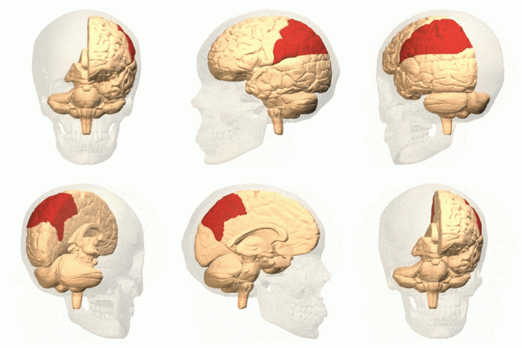
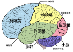
頭頂葉は大脳皮質の四つの大脳葉の一つである。中心溝の後部、外側溝(シルビウス溝)の上部、頭頂後頭溝の前方部に位置する。前頭葉とは中心溝で、側頭葉とは外側溝で、後頭葉とは内側面にある頭頂後頭溝で区切られる。頭頂葉の最前部である中心後回には一次体性感覚野がある。その後部には頭頂連合野があり、頭頂間溝によって上頭頂小葉と下頭頂小葉に分けられる。また、外側溝の中、頭頂弁蓋の内壁には二次体性感覚野がある。


解剖学的な区分
頭頂葉は大脳皮質の四つの大脳葉の一つである。頭頂葉は中心溝の後部、外側溝(シルビウス溝)の上部、頭頂後頭溝の前方部に位置する。前頭葉とは中心溝で、側頭葉とは外側溝で、後頭葉とは内側面にある頭頂後頭溝で区切られる。脳の外側面では後頭葉との間にははっきりとした脳溝がない。頭頂葉の最前部である中心後回には一次体性感覚野がある。その後部には頭頂連合野があり、頭頂間溝によって上頭頂小葉と下頭頂小葉に分けられる。また、外側溝の中、頭頂弁蓋の内壁には二次体性感覚野がある。
サルを用いた単一ニューロン活動の記録実験により、領野ごとの機能的特性が明らかにされている。本稿ではサルで得られた知見を中心に、頭頂葉の領野区分と各領野の機能的特性について簡単に紹介する。
体性感覚野
一次体性感覚野
一次体性感覚野は、3a野、3b野、2野、1野の順に分けられている。一次体性感覚野は視床の腹側基底核群からの入力を受ける。この入力は主に3野に入る。3a野には主に関節や筋などの深部受容器からの情報が入力する。3b野には皮膚受容器からの情報が多い[1]。3a、3b野は1野、2野と皮質間結合で結ばれている。1野、2野は前方の運動野、後方の頭頂連合野に投射する。一次体性感覚野は中心溝を挟んで向かい合う運動野と対称な体部位再現を持つ。この体部位再現地図は身体の物理的な広さだけでは決まっておらず、顔や手などの高精度の触感覚が必要とされる部位に対しては広い皮質領域で表現されている。
一次体性感覚野の受容野は概して小さい。触刺激の方位に対して選択性を示すニューロンも存在するが、その場合も刺激位置に対する不変性はない[2]。手指領域においては、3野ニューロンの受容野は1本の指に限局して細かい。一方、1野と2野では2本の指にまたがるような比較的広い受容野を持つニューロンや、皮膚と深部の両方の受容器からの入力を受けるニューロンがみられる[3]。
二次体性感覚野
二次体性感覚野は外側溝を上方から覆う頭頂弁蓋の内側壁に存在し、一次体性感覚野からの入力を受ける。二次体性感覚野のニューロンの受容野は一次体性感覚野のニューロンの受容野に比べて広いが、二次体性感覚野にもおおまかな体部位局在が存在する。触刺激の方位に対する選択性と受容野内での刺激位置に対する不変性を併せ持つニューロンが存在する[4]。
ヒトを被験者にしたfMRI実験から、一次体性感覚野は物理的な刺激が与えられないと賦活しないが、二次体性感覚野は実際の刺激が与えられなくても他人が触られている映像を観察しただけで賦活することが示されている[5]。また、「皮膚に注射針が刺された」画像などを観察して、痛みが想像できるような状況に置かれた場合でも二次体性感覚野は賦活する[6]。
頭頂連合野
頭頂連合野は中心溝の後方にある一次体性感覚野、腹側前方にある二次体性感覚野を除く頭頂葉の部分である。空間認知、運動視覚、高次の体性感覚の処理、手や腕等の運動制御、言語機能等、様々な認知機能に関わる。頭頂連合野外側は頭頂間溝を境として上頭頂小葉と下頭頂小葉に分けられる。なお、ブロードマンの脳区分において、ヒトでは上頭頂小葉に5野と7野の両方が当てられているが、サルでは上頭頂小葉と下頭頂小葉がそれぞれ5野と7野とされているので、ヒトとサルの対応を考える場合には注意が必要である。
サルでは、ニューロン活動が示す性質から、頭頂連合野外側の頭頂間溝領域はLIP野(外側頭頂間溝野、lateral intraparietal area)、MIP野(内側頭頂間溝野、medial intraparietal area)、AIP野(前頭頂間溝野、anterior intraparietal area)、VIP野(腹側頭頂間溝野、ventral intraparietal area)、PIP野(後頭頂間溝野、posterior intraparietal area)、CIP野(尾側頭頂間溝野, caudal intraparietal area)などに、下頭頂小葉は7a野、7b野に区分される。また、頭頂連合野内側は、頭頂後頭溝吻側壁に沿って腹側部がPO野(parieto-occipital area)、背側部がPOa野と呼ばれている。その前方の内側面は腹側部が7m野、背側部の部分がMDP野(medial dorsal parietal area)に区分される。
5野
サルでは5野は上頭頂小葉と頭頂間溝内側壁に存在する。この領野には手や上肢への触刺激や上肢の関節角などの体性感覚に応答を示すニューロンが存在する[7]。また、頭頂間溝内側壁には体性感覚の受容野近傍で呈示された視覚刺激に対して応答を示すニューロン群が存在する。遠方にある餌を手元に寄せることができるようにサルにレーキ(熊手のような道具)を使わせると、視覚性応答が得られる空間位置がレーキの先端部や到達可能な範囲にまで拡張される。このようなニューロン群は体性感覚と視覚を統合した身体像を表現していると考えられている[8][9]。
LIP野
頭頂間溝外側壁の後方半領域を占める領野であり、多くの視覚領野から入力を受け[10]、サッカード眼球運動系の中枢である前頭眼野(the frontal eye field, FEF)や上丘(the superior colliculus, SC)に直接の神経結合がある[11][12][13][14]。LIP野には視覚性反応とサッカード関連活動を示すニューロンが多く存在する。特に、記憶誘導性サッカード課題において遅延期間中に強い活動を示すニューロンの存在は近傍の領野にない特徴である[15][16][17]。これらのニューロン活動は、注意[18][19][20]、企図・意図[21][22][23]、視覚探索[24][25][26][27]、意思決定[28] [29]、報酬予測[30]などはさまざまな認知機能に関連して変調されるので、LIP野は視覚から運動への変換過程に関わる認知行動において重要な役割を果たすと考えられている。
AIP野
頭頂間溝の外側壁前方部にある領野であり、手の運動に関わる腹側運動前野(F5)との間に双方向性の神経結合がある。AIP野には、サルが物体を手で操作する際に物体の形状に選択的に活動するニューロン群が存在し、これらのニューロン群は視覚優位型、視覚運動型、運動優位型の3つのタイプに分けられる[31]。手の運動によって物体を操作する場合、操作対象となる物体の形状に応じて手の形状を細かく調整する必要がある。AIP野は、操作対象物体の形状の情報を腹側運動前野に送ることにより、手操作運動の制御に密接に関連すると考えられている[32]。
VIP野
頭頂間溝の底部にある領野であり、視覚刺激の動きに良く応答するニューロン群が存在し、多くは動き方向に対して選択的である。顔に近い場所で動く刺激に対して良く応答するニューロンや、近づいてくる刺激や遠ざかる刺激に対して選択的に応答するニューロンも見つかる。多くのニューロンが体性感覚刺激にも応答する。体性感覚の受容野位置は主として顔か頭部であり、個々のニューロンにおいては、視覚と体性感覚の受容野位置、サイズ、及び、動き刺激に対する方向選択性が一致することが多い[33]。
CIP野
頭頂間溝外側壁の後方部にある領野であり、3次元空間内で特定の方位に傾いた棒に視覚的に反応するニューロン、および3次元空間内で特定の方位に傾いた面に視覚的に反応するニューロンが多く存在する[31]。多くのニューロンにおいて、面への反応の方位選択性は両眼視差の勾配およびテクスチャーの勾配の両方を手がかりにしている[34]。
7a野
7a野(area 7a)は下頭頂小葉の後方半領域を占める領野であり、静止した対象物を注視している時に活動する注視ニューロンが存在し、その多くは注視点位置(あるいは視線方向)・距離に対して選択的である。また、動く視標を追跡する時に活動する 追跡ニューロンも報告されている[31]。視覚探索、注意に関連したニューロン活動も報告されている[35][36]。側頭葉のIT野や前頭前野の46野と直接の神経結合があることから、LIP野などの頭頂連合野内の他の領野より情報処理の階層としてより上位に存在するとの考え方もある[37]。
7b野
7b野(area 7b)は下頭頂小葉の前方半領域を占める領野であり、体性感覚刺激と視覚刺激の両方に応答するニューロン群が存在する。腹側運動前野(F5)では、他者の動作を観察している時に活動し、さらに同じ動作を自身が行っている時にも活動するニューロン群(ミラーニューロン)の存在が報告されているが、同様の性質を持つニューロン群が7b野においても見出されている[38]。これらのニューロン群は、他人の動作の理解や模倣等に関係すると考えられている。
PO野(V6野)、POa野(V6a野)、7m野、MIP野
手動作系の到達運動に関係すると考えられており、背側運動前野と双方向性の神経結合を持つ。POa野(V6a野)には頭部座標系における対象位置をコードするニューロン群、およびサルが到達運動を行っている時に方向選択性を持って活動するニューロン群のあることが知られている[39]。
関連項目
参考文献
- ↑
Jones, E.G., & Friedman, D.P. (1982).
Projection pattern of functional components of thalamic ventrobasal complex on monkey somatosensory cortex. Journal of neurophysiology, 48(2), 521-44. [PubMed:7119861] [WorldCat] [DOI] - ↑
DiCarlo, J.J., Johnson, K.O., & Hsiao, S.S. (1998).
Structure of receptive fields in area 3b of primary somatosensory cortex in the alert monkey. The Journal of neuroscience : the official journal of the Society for Neuroscience, 18(7), 2626-45. [PubMed:9502821] [WorldCat] - ↑
Iwamura, Y., Tanaka, M., Sakamoto, M., & Hikosaka, O. (1993).
Rostrocaudal gradients in the neuronal receptive field complexity in the finger region of the alert monkey's postcentral gyrus. Experimental brain research, 92(3), 360-8. [PubMed:8454001] [WorldCat] [DOI] - ↑
Thakur, P.H., Fitzgerald, P.J., Lane, J.W., & Hsiao, S.S. (2006).
Receptive field properties of the macaque second somatosensory cortex: nonlinear mechanisms underlying the representation of orientation within a finger pad. The Journal of neuroscience : the official journal of the Society for Neuroscience, 26(52), 13567-75. [PubMed:17192440] [PMC] [WorldCat] [DOI] - ↑
Keysers, C., Wicker, B., Gazzola, V., Anton, J.L., Fogassi, L., & Gallese, V. (2004).
A touching sight: SII/PV activation during the observation and experience of touch. Neuron, 42(2), 335-46. [PubMed:15091347] [WorldCat] [DOI] - ↑
Ogino, Y., Nemoto, H., Inui, K., Saito, S., Kakigi, R., & Goto, F. (2007).
Inner experience of pain: imagination of pain while viewing images showing painful events forms subjective pain representation in human brain. Cerebral cortex (New York, N.Y. : 1991), 17(5), 1139-46. [PubMed:16855007] [WorldCat] [DOI] - ↑
Mountcastle, V.B., Lynch, J.C., Georgopoulos, A., Sakata, H., & Acuna, C. (1975).
Posterior parietal association cortex of the monkey: command functions for operations within extrapersonal space. Journal of neurophysiology, 38(4), 871-908. [PubMed:808592] [WorldCat] [DOI] - ↑
Iriki, A., Tanaka, M., & Iwamura, Y. (1996).
Coding of modified body schema during tool use by macaque postcentral neurones. Neuroreport, 7(14), 2325-30. [PubMed:8951846] [WorldCat] [DOI] - ↑
Iriki, A., Tanaka, M., Obayashi, S., & Iwamura, Y. (2001).
Self-images in the video monitor coded by monkey intraparietal neurons. Neuroscience research, 40(2), 163-73. [PubMed:11377755] [WorldCat] [DOI] - ↑
Lewis, J.W., & Van Essen, D.C. (2000).
Corticocortical connections of visual, sensorimotor, and multimodal processing areas in the parietal lobe of the macaque monkey. The Journal of comparative neurology, 428(1), 112-37. [PubMed:11058227] [WorldCat] [DOI] - ↑
Schall, J.D., Morel, A., King, D.J., & Bullier, J. (1995).
Topography of visual cortex connections with frontal eye field in macaque: convergence and segregation of processing streams. The Journal of neuroscience : the official journal of the Society for Neuroscience, 15(6), 4464-87. [PubMed:7540675] [WorldCat] - ↑
Stanton, G.B., Bruce, C.J., & Goldberg, M.E. (1995).
Topography of projections to posterior cortical areas from the macaque frontal eye fields. The Journal of comparative neurology, 353(2), 291-305. [PubMed:7745137] [WorldCat] [DOI] - ↑
Andersen, R.A., Asanuma, C., Essick, G., & Siegel, R.M. (1990).
Corticocortical connections of anatomically and physiologically defined subdivisions within the inferior parietal lobule. The Journal of comparative neurology, 296(1), 65-113. [PubMed:2358530] [WorldCat] [DOI] - ↑
Paré, M., & Wurtz, R.H. (1997).
Monkey posterior parietal cortex neurons antidromically activated from superior colliculus. Journal of neurophysiology, 78(6), 3493-7. [PubMed:9405568] [WorldCat] [DOI] - ↑
Barash, S., Bracewell, R.M., Fogassi, L., Gnadt, J.W., & Andersen, R.A. (1991).
Saccade-related activity in the lateral intraparietal area. II. Spatial properties. Journal of neurophysiology, 66(3), 1109-24. [PubMed:1753277] [WorldCat] [DOI] - ↑
Barash, S., Bracewell, R.M., Fogassi, L., Gnadt, J.W., & Andersen, R.A. (1991).
Saccade-related activity in the lateral intraparietal area. I. Temporal properties; comparison with area 7a. Journal of neurophysiology, 66(3), 1095-108. [PubMed:1753276] [WorldCat] [DOI] - ↑
Gnadt, J.W., & Andersen, R.A. (1988).
Memory related motor planning activity in posterior parietal cortex of macaque. Experimental brain research, 70(1), 216-20. [PubMed:3402565] [WorldCat] [DOI] - ↑
Bisley, J.W., & Goldberg, M.E. (2003).
Neuronal activity in the lateral intraparietal area and spatial attention. Science (New York, N.Y.), 299(5603), 81-6. [PubMed:12511644] [WorldCat] [DOI] - ↑
Colby, C.L., Duhamel, J.R., & Goldberg, M.E. (1996).
Visual, presaccadic, and cognitive activation of single neurons in monkey lateral intraparietal area. Journal of neurophysiology, 76(5), 2841-52. [PubMed:8930237] [WorldCat] [DOI] - ↑
Gottlieb, J.P., Kusunoki, M., & Goldberg, M.E. (1998).
The representation of visual salience in monkey parietal cortex. Nature, 391(6666), 481-4. [PubMed:9461214] [WorldCat] [DOI] - ↑
Bracewell, R.M., Mazzoni, P., Barash, S., & Andersen, R.A. (1996).
Motor intention activity in the macaque's lateral intraparietal area. II. Changes of motor plan. Journal of neurophysiology, 76(3), 1457-64. [PubMed:8890266] [WorldCat] [DOI] - ↑
Mazzoni, P., Bracewell, R.M., Barash, S., & Andersen, R.A. (1996).
Motor intention activity in the macaque's lateral intraparietal area. I. Dissociation of motor plan from sensory memory. Journal of neurophysiology, 76(3), 1439-56. [PubMed:8890265] [WorldCat] [DOI] - ↑
Snyder, L.H., Batista, A.P., & Andersen, R.A. (1997).
Coding of intention in the posterior parietal cortex. Nature, 386(6621), 167-70. [PubMed:9062187] [WorldCat] [DOI] - ↑
Ipata, A.E., Gee, A.L., Goldberg, M.E., & Bisley, J.W. (2006).
Activity in the lateral intraparietal area predicts the goal and latency of saccades in a free-viewing visual search task. The Journal of neuroscience : the official journal of the Society for Neuroscience, 26(14), 3656-61. [PubMed:16597719] [PMC] [WorldCat] [DOI] - ↑
Ogawa, T., & Komatsu, H. (2009).
Condition-dependent and condition-independent target selection in the macaque posterior parietal cortex. Journal of neurophysiology, 101(2), 721-36. [PubMed:19073809] [WorldCat] [DOI] - ↑
Thomas, N.W., & Paré, M. (2007).
Temporal processing of saccade targets in parietal cortex area LIP during visual search. Journal of neurophysiology, 97(1), 942-7. [PubMed:17079346] [WorldCat] [DOI] - ↑
Wardak, C., Olivier, E., & Duhamel, J.R. (2002).
Saccadic target selection deficits after lateral intraparietal area inactivation in monkeys. The Journal of neuroscience : the official journal of the Society for Neuroscience, 22(22), 9877-84. [PubMed:12427844] [PMC] [WorldCat] - ↑
Churchland, A.K., Kiani, R., & Shadlen, M.N. (2008).
Decision-making with multiple alternatives. Nature neuroscience, 11(6), 693-702. [PubMed:18488024] [PMC] [WorldCat] [DOI] - ↑
Roitman, J.D., & Shadlen, M.N. (2002).
Response of neurons in the lateral intraparietal area during a combined visual discrimination reaction time task. The Journal of neuroscience : the official journal of the Society for Neuroscience, 22(21), 9475-89. [PubMed:12417672] [PMC] [WorldCat] - ↑
Platt, M.L., & Glimcher, P.W. (1999).
Neural correlates of decision variables in parietal cortex. Nature, 400(6741), 233-8. [PubMed:10421364] [WorldCat] [DOI] - ↑ 31.0 31.1 31.2
Sakata, H., Taira, M., Kusunoki, M., Murata, A., & Tanaka, Y. (1997).
The TINS Lecture. The parietal association cortex in depth perception and visual control of hand action. Trends in neurosciences, 20(8), 350-7. [PubMed:9246729] [WorldCat] [DOI] - ↑
Murata, A., Gallese, V., Luppino, G., Kaseda, M., & Sakata, H. (2000).
Selectivity for the shape, size, and orientation of objects for grasping in neurons of monkey parietal area AIP. Journal of neurophysiology, 83(5), 2580-601. [PubMed:10805659] [WorldCat] [DOI] - ↑
Duhamel, J.R., Colby, C.L., & Goldberg, M.E. (1998).
Ventral intraparietal area of the macaque: congruent visual and somatic response properties. Journal of neurophysiology, 79(1), 126-36. [PubMed:9425183] [WorldCat] [DOI] - ↑
Tsutsui, K., Sakata, H., Naganuma, T., & Taira, M. (2002).
Neural correlates for perception of 3D surface orientation from texture gradient. Science (New York, N.Y.), 298(5592), 409-12. [PubMed:12376700] [WorldCat] [DOI] - ↑
Constantinidis, C., & Steinmetz, M.A. (2001).
Neuronal responses in area 7a to multiple-stimulus displays: I. neurons encode the location of the salient stimulus. Cerebral cortex (New York, N.Y. : 1991), 11(7), 581-91. [PubMed:11415960] [WorldCat] [DOI] - ↑
Constantinidis, C., & Steinmetz, M.A. (2005).
Posterior parietal cortex automatically encodes the location of salient stimuli. The Journal of neuroscience : the official journal of the Society for Neuroscience, 25(1), 233-8. [PubMed:15634786] [PMC] [WorldCat] [DOI] - ↑
Andersen, R.A., Asanuma, C., Essick, G., & Siegel, R.M. (1990).
Corticocortical connections of anatomically and physiologically defined subdivisions within the inferior parietal lobule. The Journal of comparative neurology, 296(1), 65-113. [PubMed:2358530] [WorldCat] [DOI] - ↑
Rizzolatti, G., Fogassi, L., & Gallese, V. (2001).
Neurophysiological mechanisms underlying the understanding and imitation of action. Nature reviews. Neuroscience, 2(9), 661-70. [PubMed:11533734] [WorldCat] [DOI] - ↑
Galletti, C., Kutz, D.F., Gamberini, M., Breveglieri, R., & Fattori, P. (2003).
Role of the medial parieto-occipital cortex in the control of reaching and grasping movements. Experimental brain research, 153(2), 158-70. [PubMed:14517595] [WorldCat] [DOI]