運動前野
中山 義久、星 英司
東京都医学総合研究所 前頭葉機能プロジェクト
DOI:10.14931/bsd.5927 原稿受付日:2015年6月3日 原稿完成日:2015年11月25日
担当編集委員:一戸 紀孝(国立精神・神経医療研究センター 神経研究所)
英語名: premotor cortex 独:prämotorischer Cortex 仏:cortex prémoteur
運動前野は前頭葉にある高次運動関連領野の一つである。ブロードマン(Brodmann)の脳地図の6野の外側面を占め、一次運動野の前方、前頭前野の後方に位置する。運動前野は脳幹や脊髄に直接投射をしており運動の実行に関与する。さらに、感覚情報に基づく運動、運動の企画、運動の準備、他者の運動内容の理解(ミラーニューロン)等において、主要な役割を果たす。
歴史
ヒトの運動前野が傷害を受けると、麻痺は起こらないにもかかわらず、習熟した動作をうまく行えなくなるということが知られていた[1]。しかし、運動前野のみが傷害されることは稀であり、症状が運動前野自身の損傷によるのか、他の部位へ広がった損傷によるのかは明確ではなかった。Woolsey らは、1952年にサルの運動野を電気刺激することにより、体部位再現のマップを作成した[2]。しかし、彼らは一次運動野と運動前野を区別しなかった。さらに、脳の表面を電気刺激するという手法を用いたため、運動前野を刺激した場合であっても、その効果が一次運動野を介して現れていた可能性がある。
運動前野の機能に関する理解は、1977年のMollとKuypersによって大きく前進した[3]。彼らはサルと餌の間に透明のプラスチック板を設置し、その周囲に穴を開けた。サルは餌にまっすぐ手を伸ばすとプラスチック板によって餌を取ることができず、周辺に開けられた穴に手を回りこませて餌を取る必要があった。健常時のサルは穴を介して餌を取ることができたが、運動前野を含む領域を切除されたサルは餌に向かって手を伸ばすのみで、餌を取ることができなくなった。このことは、運動前野を中心とした領域が視覚情報を利用して運動を実現させる過程に関与することを示唆した。
その後、主にマカクザルを用いた研究により、運動前野の機能の詳細が明らかになってきた。細胞構築学的および機能的な特徴により、運動前野は背側部 (dorsal premotor cortex: PMd) と腹側部 (ventral premotor cortex: PMv) の2つの領域に分けられる[4] [5] [6]。
運動前野背側部

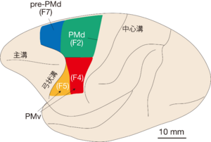
マカクザルの左半球の外側面を示す。運動前野は背側部(PMd)と腹側部(PMv)に分けられる。運動前野背側部(緑色)の前方には、前背側運動前野(pre-PMd;青色)がある。運動前野腹側部は、後方部(F4;赤色)と前方部(F5;黄色)に分けられる。
運動前野背側部(PMd)(図1、緑色の領域)の細胞の特徴として、運動の準備状態にあるときにその活動を上昇させることが挙げられる。
Wiseらは、到達するターゲットを提示し、遅延期間後のGOシグナルとともに運動を実行する課題を開発した[7]。この課題を行っている最中にサルの運動前野背側部から細胞活動を記録したところ、運動実行の際に上昇する活動(運動関連活動)に加えて、運動を指示されてからGOシグナルが提示される間に持続的に上昇する活動(準備関連活動)を多数見出した。この持続的な活動は行われる運動の内容(運動方向)を反映していたので、運動の準備状態の形成に関与すると考えられた。
さらに、運動前野背側部は、「条件つき視覚運動連合」(視覚情報に任意に連合された動作を遂行する行動)において中心的な役割を果たす。例えば、指示刺激が黄色なら左方向の運動が要求され、青色なら右方向の運動が要求されるといった場合がこれに該当する。この行動において、運動前野背側部細胞は、指示刺激そのもの(色や形など)は反映しないが、指示された動作内容をすみやかに表現し始める[8]。さらに、この行動が運動前野背側部の損傷で傷害され[9]、ヒトの脳機能画像研究はこの行動課題の遂行中に運動前野背側部の活動を同定している[10]。
これに留まらずに、複数の候補の中から一つの動作を選択する過程へ関与すること[11]、使用する手と到達するターゲットの情報を統合すること[12]、さらに、行動のゴールを運動情報へ変換する過程に関与することが[13]、運動前野背側部に見出されている。こうした一連の結果は、運動前野背側部が動作の選択や企画といった動作発現の中心となる過程に深く関与することを示す。
解剖学的研究およびヒトを対象にしたイメージング研究は運動前野背側部の前方に別の高次運動野 (前背側運動前野、図1の青色の領域) があることを示した[14] [15]。運動前野背側部は一次運動野や脊髄へ投射するが、前頭前野との連絡は限定的である。これに対し、前背側運動前野は前頭前野との連絡が強く、一次運動野や脊髄への投射はない[16] [17] [18] [19]。前背側運動前野は、次に行うべき運動の選択[20]に加えて、眼球運動と手の運動の統合[21]や、刺激属性への注意(空間的位置や形状など)[22]に関与する。また、ヒトを対象としたイメージング研究では、前背側運動前野は注意や意志決定などの認知的なプロセスに関与することが示されている[23]。
細胞構築学的な視点からは、運動前野背側部はV層の巨大錐体細胞(ベッツ細胞)の密度が一次運動野よりも低く、中程度の大きさの錐体細胞が占める[7][24][25]。また、前背側運動前野は運動前野背側部よりもはっきりした層構造が認められ、V層にはベッツ細胞は認められず、小型の錐体細胞が密集しているのが特徴である[5][15]。
また、運動前野背側部に微小電気刺激を行った場合、一次運動野で観察されるよりも高い刺激強度で閾値で電気刺激をした場合に上肢や下肢などの体部位の運動が誘発されるが、電気刺激の効果が見られない場合も多い[7][26]。一方、前背側運動前野の内側よりの部位を微小電気刺激すると急速眼球運動(サッカード)が誘発され、その部位は特に補足眼野と名付けられている[27][28]。
以上をまとめると、前背側運動前野は、前頭前野と連携して注意や意思決定などの高次機能に関与すること、それに対し、運動前野背側部は一次運動野と連携して動作の構成や実行に関与することが示唆される。運動前野背側部と前背側運動前野は相互に解剖学的な結合があるので[16]、前背側運動前野と運動前野背側部全体が連携することにより、認知情報から運動情報を生み出す様々な処理過程が実現されると考えられる。
運動前野腹側部
運動前野腹側部(PMv)細胞は、運動を行う際に視空間情報を強く表現する[29] [30] [31] [32]。さらに、運動前野腹側部はシフトプリズム順応[33]に深く関与する。こうした特徴は、運動前野背側部と同様に運動前野腹側部も視覚情報を利用した運動実行に重要であることを示唆する。運動前野腹側部は、後方部 (F4、図1の赤色の領域) と前方部 (F5、図1の黃色の領域) に分けられる[34]。F4は頭頂葉の頭頂間溝底部のVIP (ventral intraparietal area) からの入力により、F5は頭頂間溝外側壁に位置するAIP (anterior intraparietal area) や下頭頂小葉にあるPF野からの入力により特徴づけられる[35]。また、F4とF5の両者が、一次運動野と脊髄へ投射する[17] [18] [36]。
細胞構築学的には、運動前野背側部や一次運動野のIV層は萌芽的であるのに対し[37]、運動前野腹側部のIV層はより明瞭である[5]。また、Matelliらは、チトクロムオキシダーゼで染色により、F4とF5の層構造が異なることを示した。染色の濃淡によってどちらも6つの縞 (stripe) が認められるが、縞のパターンがF4とF5は異なっており、F4では4番目の薄く染まる層がIII層に含まれ5番目の濃く染まる層がほぼV層に対応するのに対し、F5は4番目と5番目の層がV層に含まれており、前頭前野と類似した特徴を有している[25]。
F4には、触覚刺激と視覚刺激の両方に応答する細胞 (bimodal neurons) が見出されている。Bimodal neuronsは、触覚刺激に応答する体部位の近傍に提示された視覚刺激に選択的に応答するという特徴がある。例えば、手の触覚刺激に応答するbimodal neuronsは、物体が手の周辺に置かれた時も強い活動を示す[38]。さらに、F4の細胞は空間的な位置情報を受け取り、それを、運動情報に変換する過程において重要な役割を果たす[33]。また、F4の細胞は、運動に関連して強い活動を示す[6]。こうした特徴は、身体周辺にある物体に手を伸ばす、あるいは、それを口で捕捉するような場合にF4が中心となることを示す。
運動前野腹側部の前方部であるF5の細胞は、物体の把持 (grasping) に関与する。Rizzolattiらは、レーズンを指先でつまむといった精密把持の時にのみ活動する細胞(grasping neuron)をF5に見出した[39]。彼らは、F5の細胞が、「つまむ」「握る」など、様々な動作の語彙 (vocabulary) を反映し、視覚情報や感覚情報によって特定の語彙が引き出されると提唱した。さらに、F5の多数の細胞は、特定の物体を見ている際に、その物体の形状選択的に活動を上昇させる[40]。こうした特徴を持つ細胞は、カノニカルニューロン (canonical neuron) と呼ばれ、最終的に運動の情報に変換するための前段階として、物体の物理的特性(大きさ、形、向きなど)を表現すると考えられる。
また、Rizzolattiらは、サルのF5の一部の細胞が、自らエサをつまむ時だけでなく、実験者がつまむ動作をするのをサルが観察している時にも、同様の活動をすることを見出した[41]。他の個体の行動を見た時に、あたかも自分がその行動を行ったかのような「鏡」のような細胞活動を示すということから、これらの神経細胞は「ミラーニューロン」と名付けられた。ミラーニューロンは、他者の運動内容の理解の基盤になっていると考えられ、さらに、他者がしていることを見て、自分がしているように感じる共感能力や意図の理解の基盤となっているという主張がなされている[42] [43]。
関連項目
参考文献
- ↑ Fulton JF.
Functional localization in the frontal lobes and cerebellum.
Clarendon Press; 1949. - ↑
WOOLSEY, C.N., SETTLAGE, P.H., MEYER, D.R., SENCER, W., PINTO HAMUY, T., & TRAVIS, A.M. (1952).
Patterns of localization in precentral and "supplementary" motor areas and their relation to the concept of a premotor area. Research publications - Association for Research in Nervous and Mental Disease, 30, 238-64. [PubMed:12983675] [WorldCat] - ↑
Moll, L., & Kuypers, H.G. (1977).
Premotor cortical ablations in monkeys: contralateral changes in visually guided reaching behavior. Science (New York, N.Y.), 198(4314), 317-9. [PubMed:410103] [WorldCat] [DOI] - ↑
Matelli, M., Luppino, G., & Rizzolatti, G. (1985).
Patterns of cytochrome oxidase activity in the frontal agranular cortex of the macaque monkey. Behavioural brain research, 18(2), 125-36. [PubMed:3006721] [WorldCat] [DOI] - ↑ 5.0 5.1 5.2
Barbas, H., & Pandya, D.N. (1987).
Architecture and frontal cortical connections of the premotor cortex (area 6) in the rhesus monkey. The Journal of comparative neurology, 256(2), 211-28. [PubMed:3558879] [WorldCat] [DOI] - ↑ 6.0 6.1
Kurata, K., & Hoffman, D.S. (1994).
Differential effects of muscimol microinjection into dorsal and ventral aspects of the premotor cortex of monkeys. Journal of neurophysiology, 71(3), 1151-64. [PubMed:8201409] [WorldCat] [DOI] - ↑ 7.0 7.1 7.2
Weinrich, M., & Wise, S.P. (1982).
The premotor cortex of the monkey. The Journal of neuroscience : the official journal of the Society for Neuroscience, 2(9), 1329-45. [PubMed:7119878] [WorldCat] - ↑
Kurata, K., & Wise, S.P. (1988).
Premotor cortex of rhesus monkeys: set-related activity during two conditional motor tasks. Experimental brain research, 69(2), 327-43. [PubMed:3345810] [WorldCat] [DOI] - ↑
Halsband, U., & Passingham, R.E. (1985).
Premotor cortex and the conditions for movement in monkeys (Macaca fascicularis). Behavioural brain research, 18(3), 269-77. [PubMed:4091963] [WorldCat] [DOI] - ↑
Toni, I., Rushworth, M.F., & Passingham, R.E. (2001).
Neural correlates of visuomotor associations. Spatial rules compared with arbitrary rules. Experimental brain research, 141(3), 359-69. [PubMed:11715080] [WorldCat] [DOI] - ↑
Cisek, P., Crammond, D.J., & Kalaska, J.F. (2003).
Neural activity in primary motor and dorsal premotor cortex in reaching tasks with the contralateral versus ipsilateral arm. Journal of neurophysiology, 89(2), 922-42. [PubMed:12574469] [WorldCat] [DOI] - ↑
Hoshi, E., & Tanji, J. (2000).
Integration of target and body-part information in the premotor cortex when planning action. Nature, 408(6811), 466-70. [PubMed:11100727] [WorldCat] [DOI] - ↑
Nakayama, Y., Yamagata, T., Tanji, J., & Hoshi, E. (2008).
Transformation of a virtual action plan into a motor plan in the premotor cortex. The Journal of neuroscience : the official journal of the Society for Neuroscience, 28(41), 10287-97. [PubMed:18842888] [PMC] [WorldCat] [DOI] - ↑
Picard, N., & Strick, P.L. (2001).
Imaging the premotor areas. Current opinion in neurobiology, 11(6), 663-72. [PubMed:11741015] [WorldCat] [DOI] - ↑ 15.0 15.1
Matelli, M., Luppino, G., & Rizzolatti, G. (1991).
Architecture of superior and mesial area 6 and the adjacent cingulate cortex in the macaque monkey. The Journal of comparative neurology, 311(4), 445-62. [PubMed:1757597] [WorldCat] [DOI] - ↑ 16.0 16.1
Luppino, G., Rozzi, S., Calzavara, R., & Matelli, M. (2003).
Prefrontal and agranular cingulate projections to the dorsal premotor areas F2 and F7 in the macaque monkey. The European journal of neuroscience, 17(3), 559-78. [PubMed:12581174] [WorldCat] [DOI] - ↑ 17.0 17.1
He, S.Q., Dum, R.P., & Strick, P.L. (1993).
Topographic organization of corticospinal projections from the frontal lobe: motor areas on the lateral surface of the hemisphere. The Journal of neuroscience : the official journal of the Society for Neuroscience, 13(3), 952-80. [PubMed:7680069] [WorldCat] - ↑ 18.0 18.1
Lu, M.T., Preston, J.B., & Strick, P.L. (1994).
Interconnections between the prefrontal cortex and the premotor areas in the frontal lobe. The Journal of comparative neurology, 341(3), 375-92. [PubMed:7515081] [WorldCat] [DOI] - ↑
Takahara, D., Inoue, K., Hirata, Y., Miyachi, S., Nambu, A., Takada, M., & Hoshi, E. (2012).
Multisynaptic projections from the ventrolateral prefrontal cortex to the dorsal premotor cortex in macaques - anatomical substrate for conditional visuomotor behavior. The European journal of neuroscience, 36(10), 3365-75. [PubMed:22882424] [WorldCat] [DOI] - ↑
di Pellegrino, G., & Wise, S.P. (1991).
A neurophysiological comparison of three distinct regions of the primate frontal lobe. Brain : a journal of neurology, 114 ( Pt 2), 951-78. [PubMed:2043959] [WorldCat] [DOI] - ↑
Mann, S.E., Thau, R., & Schiller, P.H. (1988).
Conditional task-related responses in monkey dorsomedial frontal cortex. Experimental brain research, 69(3), 460-8. [PubMed:3371430] [WorldCat] [DOI] - ↑
White, I.M., & Wise, S.P. (1999).
Rule-dependent neuronal activity in the prefrontal cortex. Experimental brain research, 126(3), 315-35. [PubMed:10382618] [WorldCat] [DOI] - ↑
Abe, M., & Hanakawa, T. (2009).
Functional coupling underlying motor and cognitive functions of the dorsal premotor cortex. Behavioural brain research, 198(1), 13-23. [PubMed:19061921] [WorldCat] [DOI] - ↑
Kurata, K., & Tanji, J. (1986).
Premotor cortex neurons in macaques: activity before distal and proximal forelimb movements. The Journal of neuroscience : the official journal of the Society for Neuroscience, 6(2), 403-11. [PubMed:3950703] [WorldCat] - ↑ 25.0 25.1
Matelli, M., Luppino, G., & Rizzolatti, G. (1985).
Patterns of cytochrome oxidase activity in the frontal agranular cortex of the macaque monkey. Behavioural brain research, 18(2), 125-36. [PubMed:3006721] [WorldCat] [DOI] - ↑
Kurata, K. (1989).
Distribution of neurons with set- and movement-related activity before hand and foot movements in the premotor cortex of rhesus monkeys. Experimental brain research, 77(2), 245-56. [PubMed:2792274] [WorldCat] [DOI] - ↑
Schlag, J., & Schlag-Rey, M. (1987).
Evidence for a supplementary eye field. Journal of neurophysiology, 57(1), 179-200. [PubMed:3559671] [WorldCat] [DOI] - ↑
Huerta, M.F., & Kaas, J.H. (1990).
Supplementary eye field as defined by intracortical microstimulation: connections in macaques. The Journal of comparative neurology, 293(2), 299-330. [PubMed:19189718] [WorldCat] [DOI] - ↑
Kubota, K., & Hamada, I. (1978).
Visual tracking and neuron activity in the post-arcuate area in monkeys. Journal de physiologie, 74(3), 297-312. [PubMed:102777] [WorldCat] - ↑
Kakei, S., Hoffman, D.S., & Strick, P.L. (2001).
Direction of action is represented in the ventral premotor cortex. Nature neuroscience, 4(10), 1020-5. [PubMed:11547338] [WorldCat] [DOI] - ↑
Hoshi, E., & Tanji, J. (2006).
Differential involvement of neurons in the dorsal and ventral premotor cortex during processing of visual signals for action planning. Journal of neurophysiology, 95(6), 3596-616. [PubMed:16495361] [WorldCat] [DOI] - ↑
Yamagata, T., Nakayama, Y., Tanji, J., & Hoshi, E. (2009).
Processing of visual signals for direct specification of motor targets and for conceptual representation of action targets in the dorsal and ventral premotor cortex. Journal of neurophysiology, 102(6), 3280-94. [PubMed:19793880] [WorldCat] [DOI] - ↑ 33.0 33.1
Kurata, K., & Hoshi, E. (1999).
Reacquisition deficits in prism adaptation after muscimol microinjection into the ventral premotor cortex of monkeys. Journal of neurophysiology, 81(4), 1927-38. [PubMed:10200227] [WorldCat] [DOI] - ↑
Rizzolatti, G., Cattaneo, L., Fabbri-Destro, M., & Rozzi, S. (2014).
Cortical mechanisms underlying the organization of goal-directed actions and mirror neuron-based action understanding. Physiological reviews, 94(2), 655-706. [PubMed:24692357] [WorldCat] [DOI] - ↑
Rizzolatti, G., Fogassi, L., & Gallese, V. (2002).
Motor and cognitive functions of the ventral premotor cortex. Current opinion in neurobiology, 12(2), 149-54. [PubMed:12015230] [WorldCat] [DOI] - ↑
Borra, E., Belmalih, A., Gerbella, M., Rozzi, S., & Luppino, G. (2010).
Projections of the hand field of the macaque ventral premotor area F5 to the brainstem and spinal cord. The Journal of comparative neurology, 518(13), 2570-91. [PubMed:20503428] [WorldCat] [DOI] - ↑
García-Cabezas, M.Á., & Barbas, H. (2014).
Area 4 has layer IV in adult primates. The European journal of neuroscience, 39(11), 1824-34. [PubMed:24735460] [PMC] [WorldCat] [DOI] - ↑
Fogassi, L., Gallese, V., Fadiga, L., Luppino, G., Matelli, M., & Rizzolatti, G. (1996).
Coding of peripersonal space in inferior premotor cortex (area F4). Journal of neurophysiology, 76(1), 141-57. [PubMed:8836215] [WorldCat] [DOI] - ↑
Rizzolatti, G., Camarda, R., Fogassi, L., Gentilucci, M., Luppino, G., & Matelli, M. (1988).
Functional organization of inferior area 6 in the macaque monkey. II. Area F5 and the control of distal movements. Experimental brain research, 71(3), 491-507. [PubMed:3416965] [WorldCat] [DOI] - ↑
Murata, A., Fadiga, L., Fogassi, L., Gallese, V., Raos, V., & Rizzolatti, G. (1997).
Object representation in the ventral premotor cortex (area F5) of the monkey. Journal of neurophysiology, 78(4), 2226-30. [PubMed:9325390] [WorldCat] [DOI] - ↑
Gallese, V., Fadiga, L., Fogassi, L., & Rizzolatti, G. (1996).
Action recognition in the premotor cortex. Brain : a journal of neurology, 119 ( Pt 2), 593-609. [PubMed:8800951] [WorldCat] [DOI] - ↑
Gallese, V., Keysers, C., & Rizzolatti, G. (2004).
A unifying view of the basis of social cognition. Trends in cognitive sciences, 8(9), 396-403. [PubMed:15350240] [WorldCat] [DOI] - ↑
Rizzolatti, G., & Craighero, L. (2004).
The mirror-neuron system. Annual review of neuroscience, 27, 169-92. [PubMed:15217330] [WorldCat] [DOI]