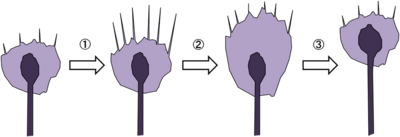
成長円錐は伸長中の神経突起の先端部に見られるアメーバ状の構造物である(図1)。19世紀にスペインの神経科学者Ramón y Cajalにより、固定染色した神経組織において神経軸索先端部に円錐状の構造が発見され、growth cone=成長円錐と名付けられた。2次元基質上で培養した場合は薄く扁平な形態をとり、多くが伸長中の神経軸索の先端に存在するが樹状突起の先端にも存在する。また、PC12細胞やN1E-115細胞のような株化細胞から伸びる神経突起様構造物の先端にも見られる。軸索の成長円錐の場合、標的神経細胞の樹状突起や組織へと到達した後は形態変化を起こしシナプス前部となる。成長円錐は極めて高い運動性を示し、細胞骨格や接着分子、膜輸送経路の制御を通じて前方へと移動し、神経突起を牽引することで伸長させる。また、成長円錐の形質膜には軸索ガイダンス因子に対する受容体が多数発現しており、軸索の成長円錐は細胞外環境に存在する軸索ガイダンス因子に応じてその運動性と進行方向を変化させ、神経軸索を正しい標的細胞へと投射させる。
上述したとおり、成長円錐は軸索ガイダンス因子に対する応答性(反応の有無、誘引-反発の方向)を場所や時期により変化させる。また、成長円錐は生体内で様々な軸索ガイダンス因子のシグナルを受容しており、成長円錐の旋回方向は多様な細胞内シグナル伝達経路の複雑なクロストークの結果決定されると考えられる。近年、成長円錐の旋回方向(誘引 or 反発)を決定する分子メカニズムの理解が急速に進んでおり、以下に旋回方向を規定する因子について概説する。
↑Schaefer, A.W., Kabir, N., & Forscher, P. (2002).
Filopodia and actin arcs guide the assembly and transport of two populations of microtubules with unique dynamic parameters in neuronal growth cones. The Journal of cell biology, 158(1), 139-52.
[PubMed:12105186]
[PMC]
[WorldCat]
[DOI]
↑Zhang, X.F., Schaefer, A.W., Burnette, D.T., Schoonderwoert, V.T., & Forscher, P. (2003).
Rho-dependent contractile responses in the neuronal growth cone are independent of classical peripheral retrograde actin flow. Neuron, 40(5), 931-44.
[PubMed:14659092]
[WorldCat]
[DOI]
↑Lee, S., & Kolodziej, P.A. (2002).
Short Stop provides an essential link between F-actin and microtubules during axon extension. Development (Cambridge, England), 129(5), 1195-204.
[PubMed:11874915]
[WorldCat]
↑Rothenberg, M.E., Rogers, S.L., Vale, R.D., Jan, L.Y., & Jan, Y.N. (2003).
Drosophila pod-1 crosslinks both actin and microtubules and controls the targeting of axons. Neuron, 39(5), 779-91.
[PubMed:12948445]
[WorldCat]
[DOI]
↑Jay, D.G. (2000).
The clutch hypothesis revisited: ascribing the roles of actin-associated proteins in filopodial protrusion in the nerve growth cone. Journal of neurobiology, 44(2), 114-25.
[PubMed:10934316]
[WorldCat]
↑Diefenbach, T.J., Latham, V.M., Yimlamai, D., Liu, C.A., Herman, I.M., & Jay, D.G. (2002).
Myosin 1c and myosin IIB serve opposing roles in lamellipodial dynamics of the neuronal growth cone. The Journal of cell biology, 158(7), 1207-17.
[PubMed:12356865]
[PMC]
[WorldCat]
[DOI]
↑Medeiros, N.A., Burnette, D.T., & Forscher, P. (2006).
Myosin II functions in actin-bundle turnover in neuronal growth cones. Nature cell biology, 8(3), 215-26.
[PubMed:16501565]
[WorldCat]
[DOI]
↑Shimada, T., Toriyama, M., Uemura, K., Kamiguchi, H., Sugiura, T., Watanabe, N., & Inagaki, N. (2008).
Shootin1 interacts with actin retrograde flow and L1-CAM to promote axon outgrowth. The Journal of cell biology, 181(5), 817-29.
[PubMed:18519736]
[PMC]
[WorldCat]
[DOI]
↑Nishimura, K., Yoshihara, F., Tojima, T., Ooashi, N., Yoon, W., Mikoshiba, K., ..., & Kamiguchi, H. (2003).
L1-dependent neuritogenesis involves ankyrinB that mediates L1-CAM coupling with retrograde actin flow. The Journal of cell biology, 163(5), 1077-88.
[PubMed:14657231]
[PMC]
[WorldCat]
[DOI]
↑Marsick, B.M., San Miguel-Ruiz, J.E., & Letourneau, P.C. (2012).
Activation of ezrin/radixin/moesin mediates attractive growth cone guidance through regulation of growth cone actin and adhesion receptors. The Journal of neuroscience : the official journal of the Society for Neuroscience, 32(1), 282-96.
[PubMed:22219290]
[PMC]
[WorldCat]
[DOI]
↑Kamiguchi, H., & Lemmon, V. (2000).
Recycling of the cell adhesion molecule L1 in axonal growth cones. The Journal of neuroscience : the official journal of the Society for Neuroscience, 20(10), 3676-86.
[PubMed:10804209]
[PMC]
[WorldCat]
↑Kamiguchi, H., & Yoshihara, F. (2001).
The role of endocytic l1 trafficking in polarized adhesion and migration of nerve growth cones. The Journal of neuroscience : the official journal of the Society for Neuroscience, 21(23), 9194-203.
[PubMed:11717353]
[PMC]
[WorldCat]
↑Dickson, B.J. (2002).
Molecular mechanisms of axon guidance. Science (New York, N.Y.), 298(5600), 1959-64.
[PubMed:12471249]
[WorldCat]
[DOI]
↑Song, H.J., & Poo, M.M. (1999).
Signal transduction underlying growth cone guidance by diffusible factors. Current opinion in neurobiology, 9(3), 355-63.
[PubMed:10395576]
[WorldCat]
[DOI]
↑Serafini, T., Colamarino, S.A., Leonardo, E.D., Wang, H., Beddington, R., Skarnes, W.C., & Tessier-Lavigne, M. (1996).
Netrin-1 is required for commissural axon guidance in the developing vertebrate nervous system. Cell, 87(6), 1001-14.
[PubMed:8978605]
[WorldCat]
[DOI]
↑Barallobre, M.J., Pascual, M., Del Río, J.A., & Soriano, E. (2005).
The Netrin family of guidance factors: emphasis on Netrin-1 signalling. Brain research. Brain research reviews, 49(1), 22-47.
[PubMed:15960985]
[DOI]
↑Hong, K., Hinck, L., Nishiyama, M., Poo, M.M., Tessier-Lavigne, M., & Stein, E. (1999).
A ligand-gated association between cytoplasmic domains of UNC5 and DCC family receptors converts netrin-induced growth cone attraction to repulsion. Cell, 97(7), 927-41.
[PubMed:10399920]
[WorldCat]
[DOI]
↑Tamagnone, L., & Comoglio, P.M. (2000).
Signalling by semaphorin receptors: cell guidance and beyond. Trends in cell biology, 10(9), 377-83.
[PubMed:10932095]
[WorldCat]
↑Taniguchi, M., Yuasa, S., Fujisawa, H., Naruse, I., Saga, S., Mishina, M., & Yagi, T. (1997).
Disruption of semaphorin III/D gene causes severe abnormality in peripheral nerve projection. Neuron, 19(3), 519-30.
[PubMed:9331345]
[WorldCat]
[DOI]
↑Dubkin, B.P., Govorunov, G.V., & Kreĭndlin, I.u.Z. (1977).
[Reconstructive operation of the vessels in ischemia of the lower extremities]. Rekonstruktivnye operatsii na sosudakh pri ishemii nizhnikh konechnosteĭ Khirurgiia, (1), 36-40.
[PubMed:846113]
[WorldCat]
↑Tear, G., Harris, R., Sutaria, S., Kilomanski, K., Goodman, C.S., & Seeger, M.A. (1996).
commissureless controls growth cone guidance across the CNS midline in Drosophila and encodes a novel membrane protein. Neuron, 16(3), 501-14.
[PubMed:8785048]
[WorldCat]
[DOI]
↑Flanagan, J.G., & Vanderhaeghen, P. (1998).
The ephrins and Eph receptors in neural development. Annual review of neuroscience, 21, 309-45.
[PubMed:9530499]
[WorldCat]
[DOI]
↑Augsburger, A., Schuchardt, A., Hoskins, S., Dodd, J., & Butler, S. (1999).
BMPs as mediators of roof plate repulsion of commissural neurons. Neuron, 24(1), 127-41.
[PubMed:10677032]
[WorldCat]
[DOI]
↑Charron, F., Stein, E., Jeong, J., McMahon, A.P., & Tessier-Lavigne, M. (2003).
The morphogen sonic hedgehog is an axonal chemoattractant that collaborates with netrin-1 in midline axon guidance. Cell, 113(1), 11-23.
[PubMed:12679031]
[WorldCat]
[DOI]
↑Huang, E.J., & Reichardt, L.F. (2001).
Neurotrophins: roles in neuronal development and function. Annual review of neuroscience, 24, 677-736.
[PubMed:11520916]
[PMC]
[WorldCat]
[DOI]
↑Gundersen, R.W., & Barrett, J.N. (1979).
Neuronal chemotaxis: chick dorsal-root axons turn toward high concentrations of nerve growth factor. Science (New York, N.Y.), 206(4422), 1079-80.
[PubMed:493992]
[WorldCat]
[DOI]
↑Tucker, K.L., Meyer, M., & Barde, Y.A. (2001).
Neurotrophins are required for nerve growth during development. Nature neuroscience, 4(1), 29-37.
[PubMed:11135642]
[WorldCat]
[DOI]
↑Lowery, L.A., & Van Vactor, D. (2009).
The trip of the tip: understanding the growth cone machinery. Nature reviews. Molecular cell biology, 10(5), 332-43.
[PubMed:19373241]
[PMC]
[WorldCat]
[DOI]
↑Myers, J.P., & Gomez, T.M. (2011).
Focal adhesion kinase promotes integrin adhesion dynamics necessary for chemotropic turning of nerve growth cones. The Journal of neuroscience : the official journal of the Society for Neuroscience, 31(38), 13585-95.
[PubMed:21940449]
[PMC]
[WorldCat]
[DOI]
↑Song, H.J., Ming, G.L., & Poo, M.M. (1997).
cAMP-induced switching in turning direction of nerve growth cones. Nature, 388(6639), 275-9.
[PubMed:9230436]
[WorldCat]
[DOI]
↑Song, H., Ming, G., He, Z., Lehmann, M., McKerracher, L., Tessier-Lavigne, M., & Poo, M. (1998).
Conversion of neuronal growth cone responses from repulsion to attraction by cyclic nucleotides. Science (New York, N.Y.), 281(5382), 1515-8.
[PubMed:9727979]
[WorldCat]
[DOI]
↑Nishiyama, M., Hoshino, A., Tsai, L., Henley, J.R., Goshima, Y., Tessier-Lavigne, M., ..., & Hong, K. (2003).
Cyclic AMP/GMP-dependent modulation of Ca2+ channels sets the polarity of nerve growth-cone turning. Nature, 423(6943), 990-5.
[PubMed:12827203]
[WorldCat]
[DOI]
↑Zheng, J.Q. (2000).
Turning of nerve growth cones induced by localized increases in intracellular calcium ions. Nature, 403(6765), 89-93.
[PubMed:10638759]
[WorldCat]
[DOI]
↑Tojima, T., Hines, J.H., Henley, J.R., & Kamiguchi, H. (2011).
Second messengers and membrane trafficking direct and organize growth cone steering. Nature reviews. Neuroscience, 12(4), 191-203.
[PubMed:21386859]
[PMC]
[WorldCat]
[DOI]
↑Ooashi, N., Futatsugi, A., Yoshihara, F., Mikoshiba, K., & Kamiguchi, H. (2005).
Cell adhesion molecules regulate Ca2+-mediated steering of growth cones via cyclic AMP and ryanodine receptor type 3. The Journal of cell biology, 170(7), 1159-67.
[PubMed:16172206]
[PMC]
[WorldCat]
[DOI]
↑Wen, Z., Guirland, C., Ming, G.L., & Zheng, J.Q. (2004).
A CaMKII/calcineurin switch controls the direction of Ca(2+)-dependent growth cone guidance. Neuron, 43(6), 835-46.
[PubMed:15363394]
[WorldCat]
[DOI]
↑Tojima, T., Hines, J.H., Henley, J.R., & Kamiguchi, H. (2011).
Second messengers and membrane trafficking direct and organize growth cone steering. Nature reviews. Neuroscience, 12(4), 191-203.
[PubMed:21386859]
[PMC]
[WorldCat]
[DOI]